
Mechanistic and Functional Evidence
Eleven therapeutic areas, twenty-two studies. Choose an area to see its summary and studies; open a study to read it in full.
In this section
- 01In Vitro Functional Studies
- 02Animal Pharmacology Studies
- 03Translational Mechanistic Evidence
Scientific validation of Dabur Chyawanprash through multi-domain preclinical evaluation
To provide scientific substantiation for the traditional Rasayana claims of Chyawanprash, Althea DRF Lifesciences undertook a comprehensive preclinical research program using validated cell-based and animal models. The objective was to systematically evaluate the efficacy and safety of Dabur Chyawanprash (DCP) across multiple physiological systems associated with immunity, resilience, healthy ageing, and overall wellbeing.
Recognizing that Chyawanprash is a complex polyherbal formulation containing diverse phytochemicals that act synergistically, the evaluation strategy was designed to examine its biological effects at functional, cellular, and organismal levels. Rather than focusing on a single therapeutic indication, studies were conducted across a broad spectrum of health domains that collectively represent the holistic benefits traditionally attributed to Rasayana therapy.
Functional cell based and cell free bioassays
Murine models under IAEC approved protocols
Mechanistic evidence linked to clinical outcomes
Read the full overview
The efficacy assessment encompassed thirteen key biological domains. These studies employed validated experimental models relevant to each physiological function and generated a comprehensive evidence base supporting the multifaceted health benefits of the formulation.
Across multiple experimental systems, Dabur Chyawanprash demonstrated significant biological activities associated with enhanced cellular protection, modulation of inflammatory responses, maintenance of tissue function, and improved physiological resilience.
Notably, several beneficial effects converged on mechanisms related to oxidative stress reduction and regulation of chronic low-grade inflammation, both of which are recognized contributors to ageing and age-associated functional decline.
These findings align closely with the Ayurvedic concept of Rasayana, which emphasizes preservation of vitality, promotion of healthy ageing, and enhancement of the body’s adaptive capacity.
Subsequent mechanistic investigations, including receptor-based profiling approaches such as GPCR screening, were undertaken to further understand the molecular pathways underlying the observed functional effects. These studies suggested modulation of signaling networks associated primarily with inflammaging, cellular stress adaptation, and maintenance of physiological homeostasis.
The integrated evidence supports the view that the benefits of Dabur Chyawanprash arise from coordinated actions across multiple biological pathways rather than through a single pharmacological mechanism. Such a systems-level mode of action is consistent with the holistic principles of Ayurveda and highlights the value of evaluating traditional formulations through multidimensional scientific approaches.
Functional studies organized by therapeutic domains
Choose an areaThe efficacy assessment frameworkTraditional Rasayana claim → validated preclinical models → multi-domain evidence base.
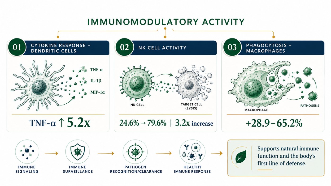 01Immune defence5.2× TNF-α3 studies
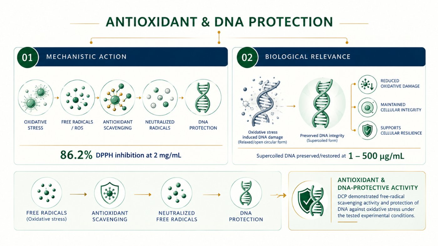 02Antioxidant and DNA protection86.2% DPPH inhibition2 studies
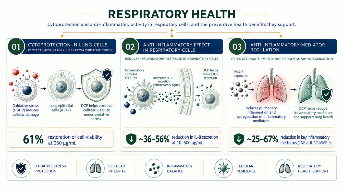 03Respiratory wellness61% Cell viability3 studies
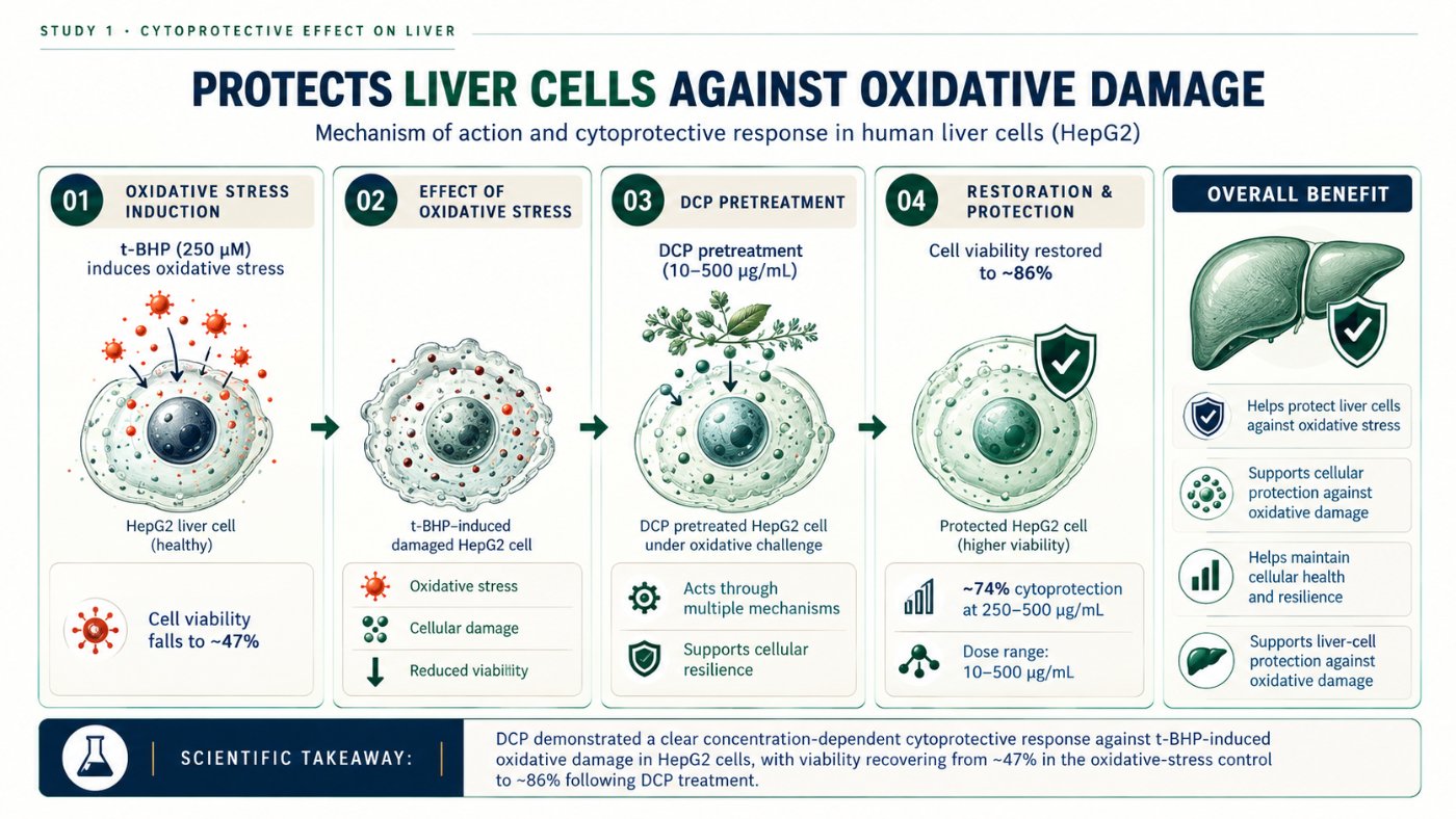 04Liver health~47 → ~86% Cell viability1 study
 05Energy and stamina2.2× ATP · 1 µg/mL1 study
 06Muscle cell resilience47% Viability · t-BHP2 studies
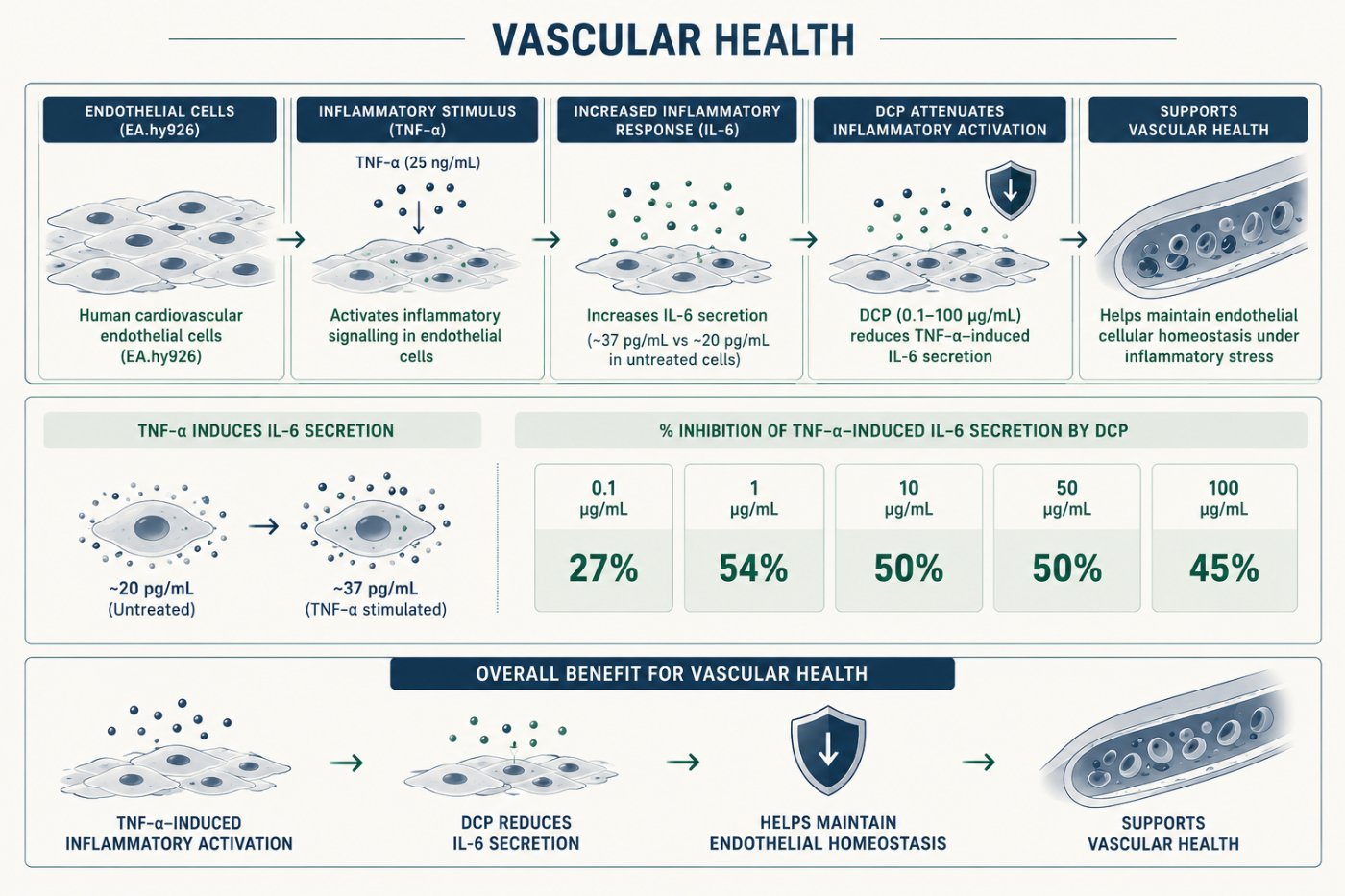 07Vascular health54% IL-6 · 1 µg/mL1 study
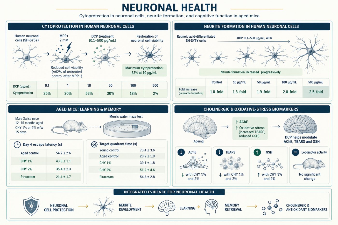 08Neuronal health53% Cytoprotection3 studies
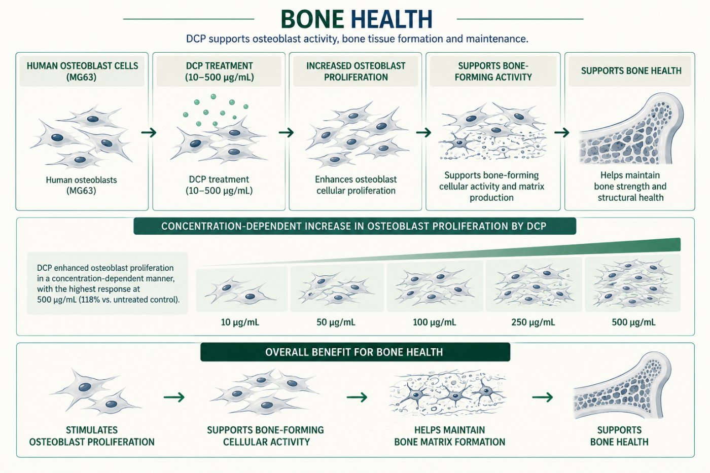 09Bone health118% Proliferation1 study
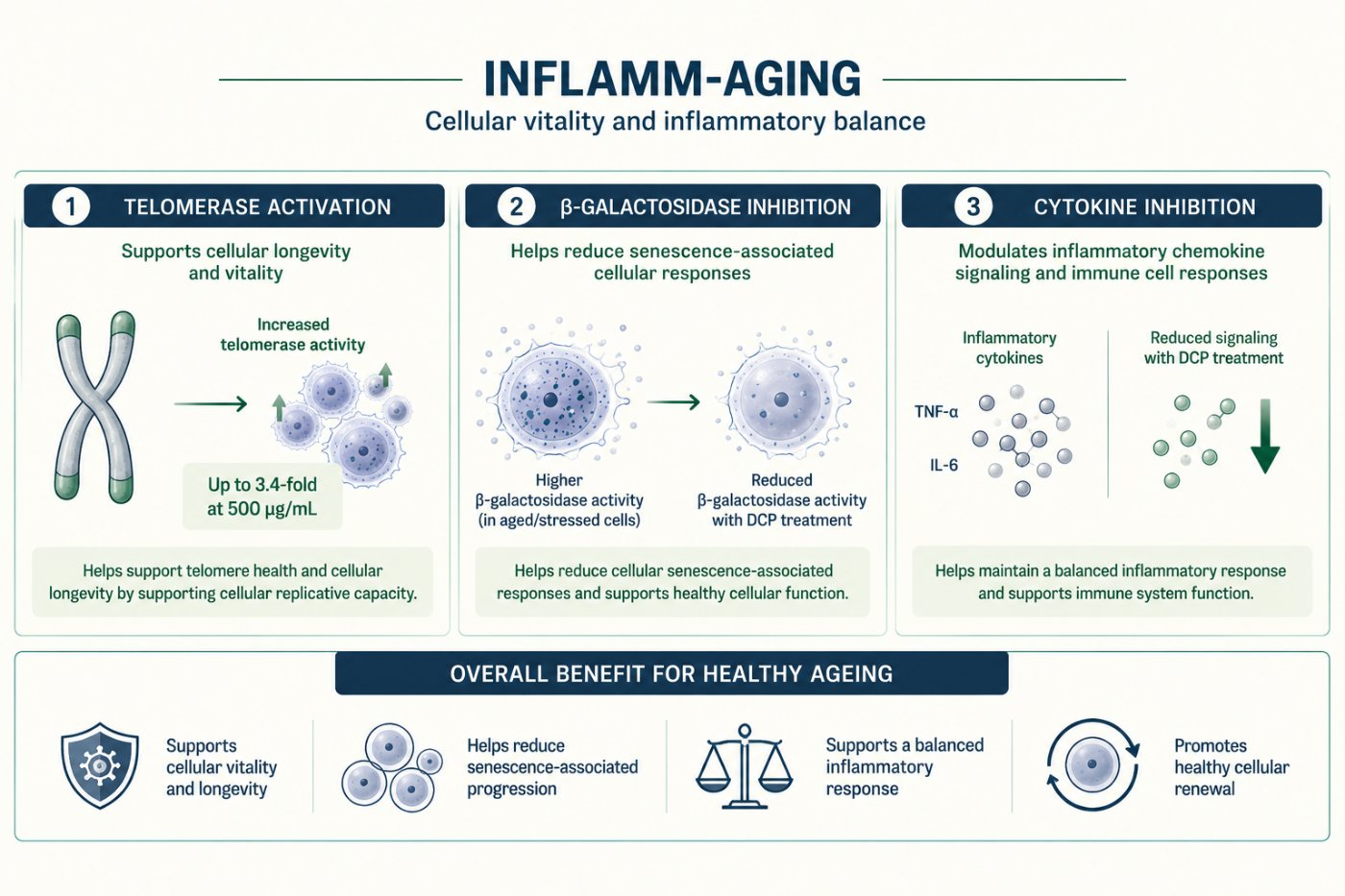 10Cellular resilience and inflammaging3.4× Telomerase3 studies
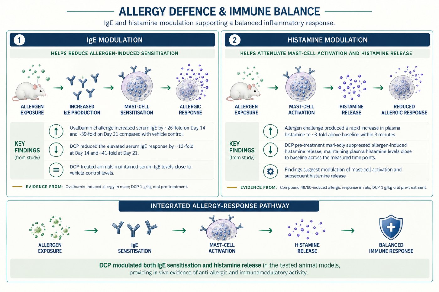 11Allergy defence~12-fold Serum IgE · Day 142 studies
Fifteen measured endpoints, read at a glance
Each panel shows one endpoint against its untreated control. Values are drawn from the studies in the eleven areas below.
2.2×
ATP · PBMCsEnergy and cellular vitality
3.2×
NK-cell activityInnate immune surveillance
5.2×
TNF-α · dendriticImmune-cell signalling
3.4×
Telomerase ActivationCellular ageing pathway
2.5×
Neurite formationNeuronal connectivity
3.3×
MIP-1α · dendriticChemokine signalling
86%
Free-radical scavengingAntioxidant protection
77%
Muscle-cell viabilityCellular resilience
74%
Liver-cell viabilityHepatocellular protection
71%
β-galactosidase InhibitionSenescence marker
65.2%
PhagocytosisMacrophage function
61%
Lung-cell viabilityRespiratory protection
56%
IL-8 inhibitionAirway inflammation
54%
IL-6 inhibitionEndothelial inflammation
+118%
Osteoblast proliferationBone-cell activity
DCP · GPCR profiling and functional validation
Experimental workflow
- 01Polyherbal master blend DCP
- 02Preparation of DCP extract
- 03GPCR screening · TansoBio reporter platform
- 04Functional assays across therapeutic areas
- 05Integrated mechanistic insight and therapeutic relevance
Overall study design
- 01Systematic GPCR profiling in agonist and antagonist modes
- 02Functional validation across eleven therapeutic areas
- 03Multi-target, multi-pathway mechanistic insight
- 04Evidence for the holistic health benefits of DCP
I · GPCR profiling, high-throughput reporter assays
Engineered HeLa cells individually expressing human GPCRs with reporter readouts. Agonist or antagonist activity measured as percentage modulation against untreated control.
HTR1AHTR2AHTR2CDRD2GABBR1/2ADRB2ADRB3MC4RFFAR1GLP1RGIPRADORA2AOXTRS1PR1
CCR2CCR5CXCR3CXCR4C5AR1AGTR1
Output. Percentage modulation, agonist and antagonist. Concentration–response curves where applicable. Benchmarking against positive controls.
II · Functional assays across multiple therapeutic areas
Immunomodulation
Immune cell activation, cytokine balance
Oxidative stress defence & fatigue support
DPPH free-radical scavenging, DNA protection
Respiratory health
Cytoprotection, anti-inflammatory
Hepatoprotection
Cytoprotection
Energy and stamina enhancement
ATP level enhancement
Muscle strength and protection
Mitochondrial potential, muscle strengthening, oxidative stress (tBHP, CoCl₂)
Cardiovascular health
Anti-inflammatory
Cognitive health
Neurite assay, neuroprotection
Bone health
Proliferation, alkaline phosphatase enhancement
Inflammaging & cellular vitality
Telomerase activity, β-galactosidase activity
Anti-allergic potential
Histamine inhibition and IgE modulation
Output. Percentage protection, activation, inhibition or enhancement. Dose–response wherever applicable.
III · Integrated data analysis and mechanistic insight
- 01GPCR signalling modulation: primary targets engagement
- 02Downstream pathways: multi-pathway effects
- 03Functional outcomes: cellular and physiological enhancement
- 04Therapeutic relevance: multi-benefit potential
Holistic mechanistic insight. Multi-target pathway modulation. Cellular protection and system-wide benefits.
Expected outcomes
- Identification of key GPCR targets modulated by DCP
- Elucidation of multi-pathway mechanisms of action
- Validation through robust functional bioassays
- Scientific rationale for broad therapeutic potential
- Scientific evidence for the holistic health benefits of DCP
A systems pharmacology approach integrating GPCR profiling with functional assays to decode the multi-target, multi-benefit potential of DCP.
Of the twenty receptors in the profiling panel, the CXCR3 result is the one reported in this review: 52% inhibition in a CXCR3-overexpressing HeLa cell model (10 · Cellular resilience and inflammaging). The workflow above sets out the design of the wider programme.
Three routes to a stronger immune response
Decoding Innate Modulation – From Cellular Mechanisms to Translational Benefits
Cytokine secretion by dendritic cells, natural killer cell cytotoxicity and macrophage phagocytosis, measured in three independent immune cell assays.
Model systemMouse BMDCs, splenic NK cells and RAW 264.7 macrophages, read against untreated control
3 studies in this area
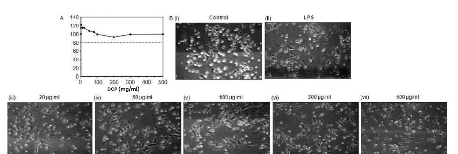 Study 1Cytokine Release StudyTo evaluate the immunostimulatory activity of Dabur Chyawanprash via cytokine release by Bone Marrow derived Dendritic Cells (BMDCs)
 Study 2NK Cell ActivityTo evaluate the immunostimulatory activity of DCP via enhanced Natural Killer (NK) cells activity.
 Study 3PhagocytosisTo evaluate the immunostimulatory activity of DCP via enhanced phagocytosis in macrophages
Mechanism
- 01DCP bioactives reach immune cells
- 02Activation of dendritic, NK and macrophage populations
- 03Cytokine release and effector function
- 04Coordinated innate immune response
Measured readouts
5.2×
Dendritic cells at 500 µg/mL (P < 0.01)
2.2×
Dendritic cell pro-inflammatory signal
3.3×
At 500 µg/mL (2.9× at 300 µg/mL)
3.2×
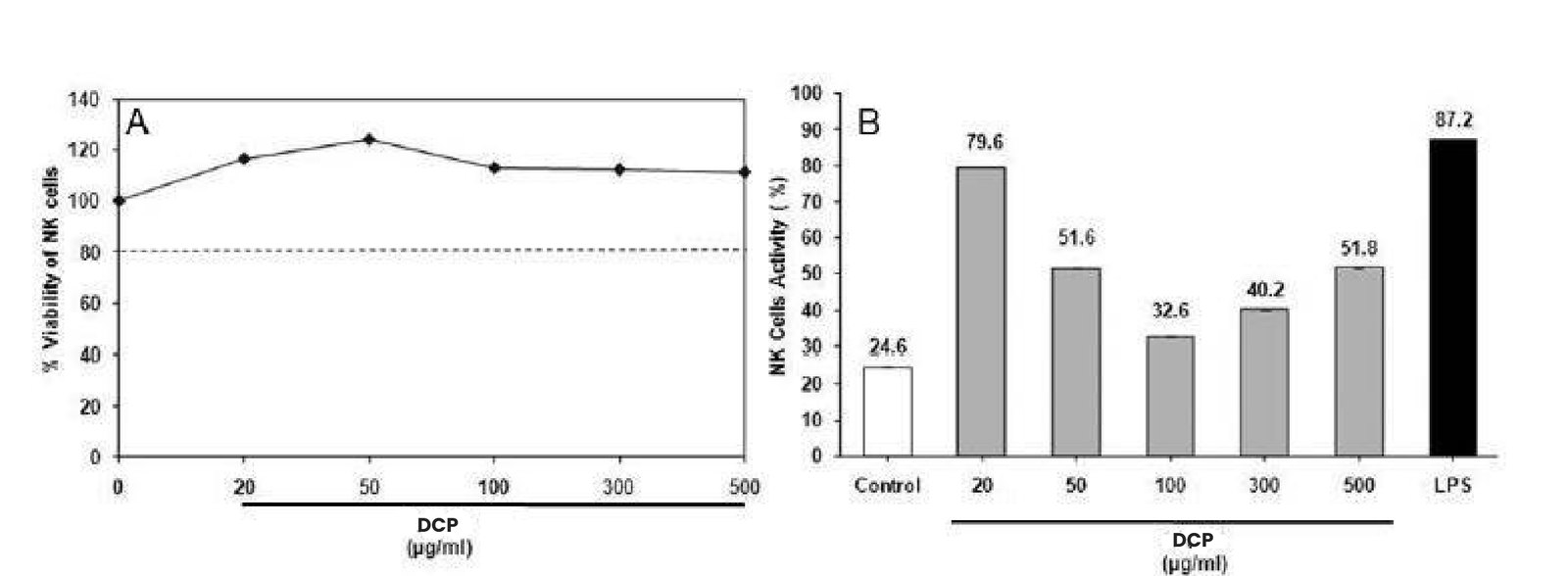
24.6% to 79.6% target lysis at 20 µg/mL
+28.9–65.2%
Zymosan uptake, 50–500 µg/mL (P < 0.01)
≥80%
No cytotoxicity across 0.1–500 µg/mL
Cellular benefits
- Enhanced immune signalling and cell communication
- Improved recruitment and activation of immune cells
- Stronger immune surveillance and pathogen control
- Supports initiation of the adaptive immune response
Interpretation
DCP acts on all three arms of innate immunity in parallel, raising signalling capacity, cytotoxic surveillance and particle clearance rather than a single isolated marker.
Supports natural immune function and strengthens the body's first line of defence.
Scientific interpretation for consumer benefit claim
Collectively, the enhanced secretion of immune signaling molecules, increased NK cell cytotoxic activity, and improved macrophage phagocytosis demonstrate that DCP exerts broad immunostimulatory effects on key components of the innate immune system. Activation of these immune functions supports efficient immune cell communication, pathogen recognition and clearance, immune surveillance, and antigen processing, which are essential for mounting an effective early immune response. By strengthening the body’s first line of defence and facilitating the transition to adaptive immunity, these findings suggest that DCP help support natural immune function, promotes immune resilience, and contributes to maintaining overall immune health.
Below consumer claims can be made:
- Supports a healthy immune system.
- Helps strengthen the body’s natural immune defences.
- Supports innate immune function, the body’s first line of defence.
- Helps the immune system respond efficiently to everyday challenges.
- Supports healthy immune cell activity.
- Promotes natural immune resilience.
- Helps maintain overall immune health and wellness.
- Supports the body’s natural defence mechanisms.
The collective findings from the in vitro immunological investigations provide a strong scientific basis for the 3X Immunity claim featured on the DCP product label. These data sets demonstrate that DCP supports multiple functional pillars of innate immunity, providing a robust scientific rationale for the 3X Immunity positioning. The claim reflects the formulation’s ability to support three key immune defence mechanisms that contribute to maintaining healthy immune function and overall immune resilience.
The mechanistic in vitro findings were consistent with the clinical outcomes, which demonstrated a 2–4-fold improvement in immune-related parameters. The concordance between the mechanistic and clinical evidence supports the biological plausibility of DCP’s immunomodulatory effects and provides a stronger scientific foundation for the 3X Immunity claim.
Tnf-
5.2-fold increase at 500 μg/ml
Nk cell activity
24.6% to 79.6%, 3.2-fold
Phagocytosis
28.9 to 65.2% increased internalization
DRF in-house immune cell assays.
Cytokine Release Study
To evaluate the immunostimulatory activity of Dabur Chyawanprash via cytokine release by Bone Marrow derived Dendritic Cells (BMDCs)
Bone Marrow derived Dendritic Cells (BMDCs) were used. Specific pathogen-free male C57BL/6 mice (20-25 g, 8–10 weeks) were obtained from National Centre for Laboratory Animal Sciences (NCLAS), National Institute of Nutrition (NIN), Hyderabad, India, and were kept in the in-house animal facility maintained at 22±3 °C and 55±15% relative humidity with 12 h L:D cycle. They were given autoclaved pelleted feed and filtered drinking water ad libitum. All experiments involving the mice were performed under the protocols approved by the Institutional Animal Ethics Committee (IAEC).
Methodology
Generation of DC cultures from bone marrow
DCs were generated from murine bone marrow. Femurs were excised from C57BL/6 mice and bone marrow was harvested by gently flushing the femur with HBSS using a 23-gauge needle.
Cells were cultured in 90-mm culture petri dishes (2 × 10⁶ cells/10 ml) with growth medium (RPMI 1640 medium supplemented with 10% FBS, 100U/ml penicillin, 100 μg/ml streptomycin and 20 ng/ml rmGMCSF) for 6 days at 37 °C in a humidified 5% CO₂ atmosphere.
On day 3, cells were replenished with additional 10 ml of growth medium containing rmGMCSF. Semi-adherent, immature DCs were harvested on day 6 by gentle pipetting and used in experiments. Viable cells were counted using trypan blue exclusion method.
Estimation of cytokines (TNF-α, IL-1-βand mip-1-α) in dcs
Effect of DCP on viability of DCs was assessed after 24 h of treatment.
DCP was dissolved in DMSO to make 200mg/ml stock concentration which was serially diluted to achieve final concentrations in cells.
DCs (50,000 cells/well) were plated in 96-well culture plates and treated with DCP in the concentration range of 0.1-500 μg/ml in triplicates. The cytotoxic effect of DCP on DCs was determined after 24 h of incubation by MTT assay. Concentrations of DCP that resulted in >80% viability of DCs were selected for subsequent cytokine analysis studies.
For estimation of cytokines activity, day-6 DCs (0.16 × 10⁶ cells/well in 24-well culture plate) were treated with DCP at selected non-cytotoxic concentrations, 20-500 μg/ml in triplicates. DCs treated with 0.25% DMSO were included as control cells.
After 24 h of incubation, cell-free culture supernatants were analyzed for secreted levels of TNF-α, IL-1-β and MIP-1-α by ELISA. LPS (E. coli) (10 ng/ml) was included as a positive control.
Key biomarkers
TNF-α (Tumor Necrosis Factor-α): Key pro-inflammatory cytokine; indicates activation of innate immune responses and inflammation.
IL-1β (Interleukin-1β): Major pro-inflammatory cytokine involved in inflammasome activation, fever, and immune cell activation.
MIP-1α (CCL3): Chemokine that recruits monocytes, macrophages, T cells, and NK cells to sites of infection or inflammation, reflecting immune cell trafficking.
Results
Dendritic cells showed ≥ 80% viability in the concentration range of 0.1-500 μg/ml for 24 h. DCP also induced remarkable changes in the morphology of DCs. Control DCs showed formation of numerous colonies with semiadherent cells in the suspension. Some cells adhering to surface were also seen at the site of colony formation. At a concentration of 20-500 μg/ml DCP causes a dose dependent maturation in DCs. This effect was evident by increased number of adhered and branched cells in comparison with semi-adherent cells. Maturation of DCs was also observed upon treatment with LPS (10 ng/ml), which was used as a positive control.
Stimulatory effects of DCP on DCs activity were determined by estimating the levels of cytokines secreted in culture supernatants using ELISA. DCP at 20-500 μg/ml for 24 h resulted in increased secretion of TNF-α, IL-1-β and MIP-1-α as compared to control DCs. TNF-α recorded a 5.2 fold significant increase (p <0.01) at 500 μg/ml followed by 2.2-fold increase of IL-1β. Levels of MIP-1α secreted into extracellular medium also showed 2.9 and 3.3 fold increase at 300 μg/ml and 500 μg/ml, respectively (p <0.01). With the positive control LPS (10 ng/ml) also, DCs showed significantly (p <0.01) increased secretion of cytokines, TNF-α, IL-1β and MIP-1α.
Scientific interpretation
DCP exhibited potent immunostimulatory activity by activating dendritic cells, as evidenced by the dose-dependent increase in TNF-α, IL-1β, and MIP-1α secretion. Elevated TNF-α and IL-1β indicate activation of innate immune and inflammatory signaling pathways, promoting dendritic cell maturation and antigen presentation, while increased MIP-1α suggests enhanced recruitment of immune cells such as monocytes, macrophages, NK cells, and T lymphocytes.
NK Cell Activity
To evaluate the immunostimulatory activity of DCP via enhanced Natural Killer (NK) cells activity.
Splenocytes derived from C57BL/6 mice were used as effector NK cells population. Mouse lymphoma cells (YAC-1) were used as target cells. Specific pathogen-free male C57BL/6 mice (20-25 g, 8–10 weeks) were obtained from National Centre for Laboratory Animal Sciences (NCLAS), National Institute of Nutrition (NIN), Hyderabad, India, and were kept in the in-house animal facility maintained at 22±3 °C and 55±15% relative humidity with 12 h L:D cycle. They were given autoclaved pelleted feed and filtered drinking water ad libitum. All experiments involving the mice were performed under the protocols approved by the Institutional Animal Ethics Committee (IAEC).
Methodology
Spleens were removed from C57BL/6 mice and a single cell suspension was prepared. Erythrocytes in the splenocytes were removed by treatment with lysis buffer (0.15 M NH₄Cl, 0.01 M NaHCO₃ and 0.1 mM Na₂EDTA, pH 7.4) for 5 min.
Cells were washed in RPMI-1640 medium and used as NK cell population. Mouse lymphoma cell line (YAC-1) (sensitive to NK cells) was maintained in RPMI-1640 supplemented with 10% FBS, Penicillin (100 U/ml), Streptomycin (100 μg/ml) at 37 °C, 5% CO₂ and 95% humidity and used as a target cell population.
To assess the effects of DCP on viability, splenocytes were seeded at a density of 0.5 × 10⁶ cells/well in 96-well culture plates and then treated with DCP (20-500 μg/ml) in triplicates. After 24 h of incubation, cytotoxic effect was determined by MTT assay.
For NK cell activity, splenocytes (0.5 × 10⁶ cells/well in 96-well culture plate) were treated with DCP at non-cytotoxic concentrations in the range 20-500 μg/ml (in triplicates) for 24 h. YAC-1 cells (5000 cells/well) were co-incubated with NK-cells pre-treated with DCP (Effector: Target ratio of 100:1). NK cells alone (treated with 0.25% DMSO) (E) and YAC-1 cells alone (T) were also incubated as controls. After 4 h of incubation at 37 °C in a CO₂ incubator, NK cells mediated lysis of YAC-1 cells was determined using MTT assay. LPS (E. Coli. 100 ng/ml) was used as a positive control.
Percentage of NK cell activity was calculated as: NKA% = [1-{O.D. - O.D. }] / O.D. × 100
Key biomarkers
NK cells activity indicates enhanced innate immune surveillance against viral infections.
Results
NK cells on treatment with DCP (20-500 μg/ml) for 24 h did not show any significant loss of cell viability.
To further evaluate the immunostimulatory activity, NK cells were first treated with DCP for 24 h and then co-cultured with YAC-1 cells at E:T ratio of 100:1. DCP, at the concentration range of 20-500 μg/ml, stimulated NK cell activity.
A basal level of 24.6% NK cell activity was observed in control cells, which increased up to a maximum of 79.6% (by 3.2-fold) at 20 μg/ml of DCP. Positive control LPS (100 ng/ml) also enhanced NK cell activity to 87.2% (by 3.5-fold).
Scientific interpretation
DCP significantly enhanced NK cell cytotoxic activity, increasing target cell lysis from a basal level of 24.6% to 79.6% (3.2-fold), demonstrating potent stimulation of innate immune effector function. The response, comparable to that of the positive control LPS, suggests that DCP activates NK cells, thereby enhancing their ability to recognize and eliminate virus-infected and transformed cells. Increased NK cell activity also supports improved immune surveillance and cytokine-mediated crosstalk with dendritic cells and other immune cells, indicating that DCP possesses strong immunostimulatory potential by reinforcing early innate immune defence and facilitating the development of adaptive immune responses.
Phagocytosis
To evaluate the immunostimulatory activity of DCP via enhanced phagocytosis in macrophages
Mouse macrophage cell line (RAW264.7) was procured from the National Centre For Cell Science (NCCS, Pune, India) and grown in DMEM supplemented with 10% FBS, 100 U/ml penicillin and 100 μg/ml streptomycin at 37 °C in a 5% humidified incubator with a 95% air atmosphere.
Methodology
Effect of DCP on viability of RAW264.7 cells was determined after 24 h. Cells were seeded at a density of 5 × 10⁴ cells/well in 96-well culture plates. After overnight incubation, cells were treated with DCP. (0.1-500 μg/ml) in triplicates for another 24 h.
Any cytotoxic effect of DCP on mouse macrophages (RAW 264.7) was determined by MTT assay.
For phagocytosis assay, RAW 264.7 cells were seeded at a density of 5 × 10⁴ cells/well in 96-well culture plates for 24 h. Cells were treated with DCP at non-cytotoxic concentrations, 20-500 μg/ml in triplicates for another 24 h. Cells treated with 0.25% DMSO were used as controls. LPS (E. coli.) (50 μg/ml) was used as a positive control.
Phagocytosis was estimated using CytoSelect™ 96-well phagocytosis assay kit (Zymosan, Colorimetric Format).
Key biomarkers
Enhanced phagocytic activity is a hallmark of innate immune activation and reflects improved functional capacity of macrophages and other phagocytes to recognize, engulf, and eliminate pathogens, cellular debris, and apoptotic cells.
Results
The mouse macrophages (RAW 264.7) did not show any significant loss of cellular viability with DCP at 0.1- 500 μg/ml after a time period of 24 h.
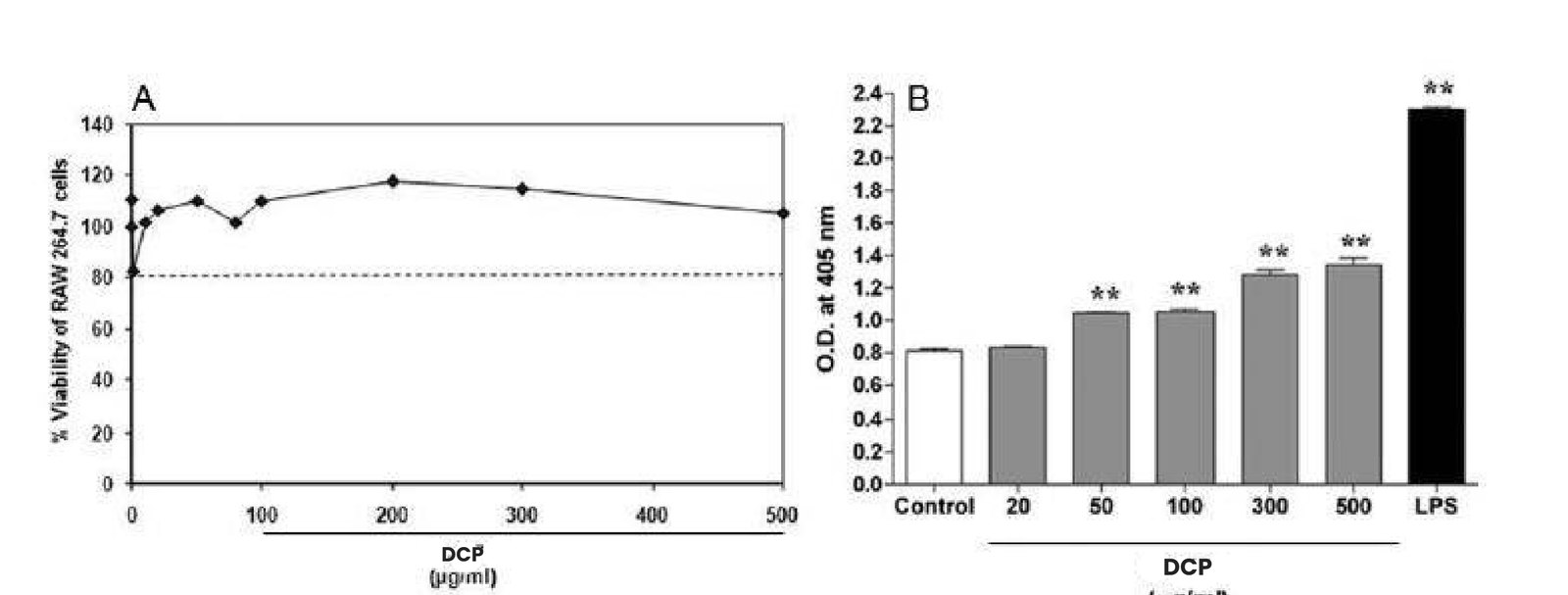
The stimulatory effect of DCP on phagocytosis was measured after 24 h of treatment by the extent of internalization of prelabeled zymosan particles using a colorimetric kit based method (Cytoselect, Cellbio Labs). Phagocytosis of macrophages showed significant increase upon treatment with DCP in the concentration range of 50-500 μg/ml (P <0.01).
Within this concentration range, DCP stimulated the extent of zymosan-particles internalization by 28.9-65.2% as compared to control RAW 264.7 cells. LPS (50 μg/ml) also significantly (P <0.01) enhanced phagocytosis of macrophages, when used as a positive control.
Scientific interpretation
DCP demonstrated immunostimulatory activity by enhancing macrophage phagocytosis without affecting cell viability, indicating functional activation rather than cytotoxicity. The increased uptake of zymosan particles reflects enhanced innate immune defence, improved pathogen clearance, and greater antigen processing, suggesting activation of macrophage-mediated immune responses and strengthening of the host’s first line of defence.
Protecting the cell’s genetic core from oxidative damage
Consolidated Evidence on Antioxidant Potential and Genomic Protection for Resilience and Fatigue Support
Free radicals generated by UV, pollution and metabolic load damage membranes, proteins and DNA.
Model systemDPPH radical scavenging assay and pBR322 plasmid DNA under H₂O₂ + UVB
2 studies in this area
 Study 1In Vitro Antioxidant AssayTo evaluate the in vitro antioxidant potential of Dabur Chyawanprash (DCP) by determining its ability to scavenge the stable free radical 2,2-diphenyl-1-picrylhydrazyl (DPPH).
 Study 2Protection of DNA Damage Against Oxidative DamageTo evaluate the in DNA damage protective potential of Dabur Chyawanprash (DCP) by determining its ability for restoration of supercoiled DNA against oxidative damage for fatigue support
Mechanism
- 01Oxidative stressors: UV, pollution, metabolic load
- 02Generation of free radicals
- 03DCP natural antioxidants neutralise radicals
- 04DNA structure and cell integrity protected
Measured readouts
86.2%
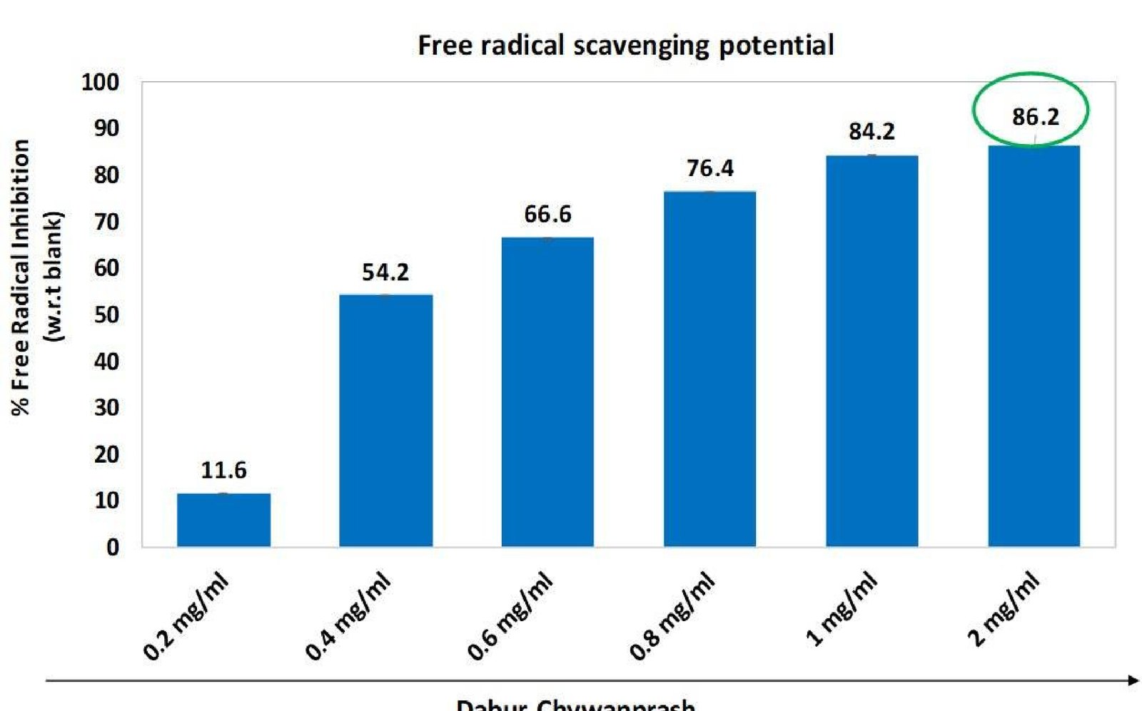
Maximum free-radical scavenging at 2 mg/mL
11.6 → 86.2%
Concentration dependent across 0.2–2 mg/mL
>84%
Sustained inhibition at ≥1 mg/mL
Restored
Native band reappears at 1–500 µg/mL
Attenuated
Less open-circular/linear shift vs H₂O₂ + UVB
No effect
Vehicle alone did not protect DNA
Cellular benefits
- Protects cells from oxidative stress
- Supports DNA integrity and genomic stability
- Reduces the cellular oxidative burden
- Supports healthy cellular ageing
Interpretation
The protection is upstream: by lowering the radical load itself, DCP limits oxidative damage before it reaches DNA, rather than repairing one endpoint after the fact.
Provides antioxidant protection and helps safeguard cellular DNA.
Scientific interpretation for consumer benefit claim
The findings provide mechanistic support for the antioxidant and cellular-protective potential of DCP. DCP preserved the supercoiled form of plasmid DNA following H O + UVB-induced oxidative damage, indicating protection against oxidative stress-related DNA damage. From a consumerhealth perspective, this suggests that DCP may help protect cellular components from oxidative stress, maintain cellular integrity and support cellular resilience during physiological stress restoration of supercoiled DNA band against UVB+oxidative stress
Below consumer claims can be made:
- Supports antioxidant protection
- Helps protect cells from oxidative stress
- Supports cellular protection against oxidative damage
- Helps protect DNA from oxidative stress-induced damage
- Supports DNA and cellular integrity
- Helps maintain cellular integrity during oxidative stress
- Supports cellular resilience during physical and metabolic stress
- Helps protect cells against oxidative stress associated with increased metabolic demand
- Supports cellular antioxidant defences for maintaining vitality
Dpph inhibition
11.6% at 0.2 mg/mL to 86.2% at 2 mg/mL
Plateau
≥1 mg/mL, inhibition exceeded 84%
Dna protection
Supercoiled band restored at 1–500 μg/mL
DRF in-house antioxidant and DNA protection assays.
In Vitro Antioxidant Assay
To evaluate the in vitro antioxidant potential of Dabur Chyawanprash (DCP) by determining its ability to scavenge the stable free radical 2,2-diphenyl-1-picrylhydrazyl (DPPH).
Cell-free biochemical antioxidant assay
Methodology
Dabur Chyawanprash (DCP) was dissolved in 80% methanol to make stock of 500 mg/ml. Further the stock solution was diluted in 80% methanol and tested against conc. ranges from 0.1mg/ml – 2mg/ml for antioxidant potential.
Dabur Chyawanprash (DCP) was evaluated for its free-radical scavenging potential at concentrations ranging from 0.1 to 2 mg/mL. The required concentrations of DCP were prepared by serial dilution of the stock solution using 80% methanol.
The prepared DCP test solutions were added to a 96-well microplate at the respective concentrations and were subsequently mixed with 250 μM DPPH (2,2-diphenyl-1-picrylhydrazyl) solution in a 1:2 ratio.
The reaction mixtures were thoroughly mixed to ensure uniform interaction between the DPPH radicals and the test samples and were incubated for 30 minutes at 37°C under protected conditions.
A reaction mixture containing DPPH and 80% methanol was used as the corresponding solvent control/blank for determination of the baseline DPPH absorbance.
Following the incubation period, the reduction in DPPH radical absorbance resulting from the free-radical scavenging activity of DCP was measured spectrophotometrically at 570 nm.
The antioxidant/free-radical scavenging activity of DCP was assessed across the tested concentration range by calculating the percentage reduction in DPPH free-radical activity relative to the control.
The concentration-dependent antioxidant response was further evaluated by plotting the percentage inhibition against the corresponding DCP concentrations, and the IC value was determined. The IC represented the concentration of DCP required to achieve 50% reduction in DPPH free-radical activity under the experimental conditions.
Key biomarkers
DPPH is a stable nitrogen-centered free radical characterized by a deep purple colour. In the presence of an antioxidant, DPPH accepts a hydrogen atom or electron from the antioxidant molecule and is converted to its reduced form, resulting in a decrease in absorbance. Thus, the reduction in DPPH absorbance following exposure to DCP is used as an indicator of its freeradical scavenging/antioxidant capacity. The magnitude of DPPH reduction is expected to increase with increasing concentrations of DCP, allowing generation of a concentration-response relationship and calculation of the IC50.
Results
Dabur Chyawanprash (DCP) demonstrated a concentration-dependent increase in DPPH free-radical scavenging activity over the tested concentration range of 0.2–2 mg/mL. The observed response indicated that DCP exhibited appreciable antioxidant/free-radical scavenging potential under the experimental conditions.
The highest tested concentration of 2 mg/mL demonstrated 86.2% free-radical inhibition, which represented the maximum activity observed in the tested concentration range.
The results therefore demonstrated that DCP possessed strong in vitro DPPH free-radical scavenging activity and exhibited a clear concentration-dependent antioxidant response, with 86.2% inhibition being achieved at 2 mg/mL under the experimental conditions.
Scientific interpretation
Dabur Chyawanprash demonstrated a pronounced concentration-dependent free-radical scavenging response, with DPPH radical inhibition increasing from 11.6% at 0.2 mg/mL to 86.2% at 2 mg/mL. The high level of radical inhibition observed at the upper concentrations indicated substantial in vitro antioxidant potential of DCP. The response approached a plateau at concentrations ≥1 mg/mL, where inhibition exceeded 84%, demonstrating consistently high free-radical scavenging activity.
Protection of DNA Damage Against Oxidative Damage
To evaluate the in DNA damage protective potential of Dabur Chyawanprash (DCP) by determining its ability for restoration of supercoiled DNA against oxidative damage for fatigue support
pBR322 Plasmid DNA from E. coli
Methodology
Dabur Chyawanprash (DCP) was dissolved in 80% methanol to make stock of 500 mg/ml. Further the stock solution was diluted in 80% methanol and tested against conc. ranges from 0.1mg/ml – 2mg/ml for antioxidant potential.
Plasmid DNA was incubated with different concentrations of Dabur Chyawanprash (DCP; 1–500 μg/mL) at 37°C for 2 hours to allow interaction between the formulation and DNA.
Following pre-incubation, the plasmid DNA was subjected to oxidative and UV-induced damage using H₂O₂ in combination with UVB irradiation (157 mJ/cm2). This treatment induces oxidative modifications and structural damage to DNA, resulting in conversion of the native supercoiled (ccc) plasmid form into more relaxed/open-circular and/or linear forms.
After damage induction, samples were mixed with 6× DNA loading dye and separated by 1.5% agarose gel electrophoresis at 90 V for 1 hour, enabling visualization and differentiation of the various structural forms of plasmid DNA.
The separated DNA bands were visualized and documented using a Bio-Rad Gel Documentation System. The relative distribution of the supercoiled, open-circular and linear DNA forms was used to assess the extent of DNA damage and protection.
Preservation or restoration of the supercoiled plasmid DNA band in DCP-treated samples compared with the H₂O₂ + UVB-damaged control indicates attenuation of oxidative/UV-induced DNA structural damage.
Thus, the assay evaluates the DNA-protective/antioxidant potential of DCP, with retention or recovery of the supercoiled DNA conformation providing evidence that DCP can help protect DNA against oxidative stress-induced structural damage.
Key biomarkers
The plasmid DNA protection assay has indirect but biologically relevant significance for fatigue and stamina, because prolonged physical activity and metabolic demand can increase the generation of reactive oxygen species (ROS). Excessive ROS can cause oxidative damage to cellular components, including DNA, proteins and lipids, potentially compromising cellular integrity and normal energy metabolism. The ability of DCP to preserve the supercoiled form of plasmid DNA following H₂O₂ + UVB-induced oxidative damage indicates that its constituents have the potential to counteract oxidative stress and protect genetic material from oxidative damage. At a broader cellular level, maintaining DNA integrity is important for normal cellular function, repair processes and sustained cellular resilience during periods of physiological stress. Therefore, the DNA-protection findings can provide mechanistic support for an antioxidant/cellular resilience pathway relevant to fatigue, particularly the concept that reducing oxidative damage may help cells better withstand stress associated with increased metabolic activity. However, DNA protection alone does not establish that DCP reduces subjective fatigue or directly improves stamina/endurance.
Results
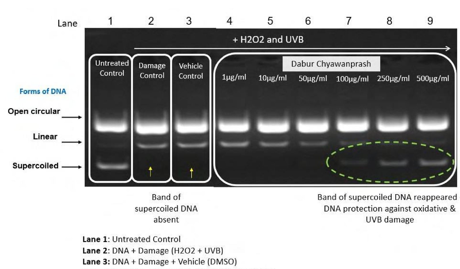
The agarose gel demonstrates clear protection of plasmid DNA by DCP against H₂O₂ + UVB-induced oxidative damage. In the untreated control (Lane 1), the characteristic supercoiled plasmid DNA band is clearly visible, representing the intact/native form of the DNA. In contrast, exposure to H₂O₂ + UVB in the damage control (Lane 2) resulted in the loss of the supercoiled DNA band, with a shift toward the open circular/linear forms, confirming substantial oxidative damage to the plasmid DNA.
The vehicle control (Lane 3) showed a similar damaged DNA pattern, indicating that the vehicle itself did not provide meaningful DNA protection. Importantly, in the presence of DCP (Lanes 4–9; 1–500 μg/mL), the supercoiled DNA band reappeared, indicating restoration/preservation of the native plasmid conformation following oxidative and UVB challenge.
The reappearance of the supercoiled form across the DCP-treated samples demonstrates that DCP attenuated H₂O₂ + UVB-induced DNA structural damage. The findings therefore provide evidence of a DNA-protective and antioxidant effect of DCP, suggesting that its constituents can help maintain DNA integrity under conditions of oxidative stress. Overall, the assay supports the potential of DCP to protect cellular genetic material against oxidative damage and enhance cellular resilience to oxidative stress.
Scientific interpretation
The findings demonstrate that DCP possesses DNA-protective activity under oxidative stress conditions. H O + UVB exposure disrupted the native supercoiled conformation of plasmid DNA, indicating oxidative/UV-induced structural damage. The reappearance of the supercoiled DNA band in DCP-treated samples (1–500 μg/mL) indicates that DCP attenuated this damage and helped preserve DNA structural integrity. Scientifically, this suggests that DCP may provide antioxidant-mediated protection against oxidative damage to cellular genetic material, thereby supporting overall cellular resilience and maintenance of cellular integrity under oxidative stress.
Defending the lining of the airway
Substantiated by Oxidative Damage Protection, Anti-inflammatory Evidence in Lung cells
The airway epithelium is the first surface exposed to airborne irritants and pathogens. DCP protects lung cells from oxidative stress and moderates the local inflammatory response.
Model systemA549 lung epithelial cells; PM2.5-exposed Balb/c mice, DCP 500 and 2000 mg/kg
3 studies in this area
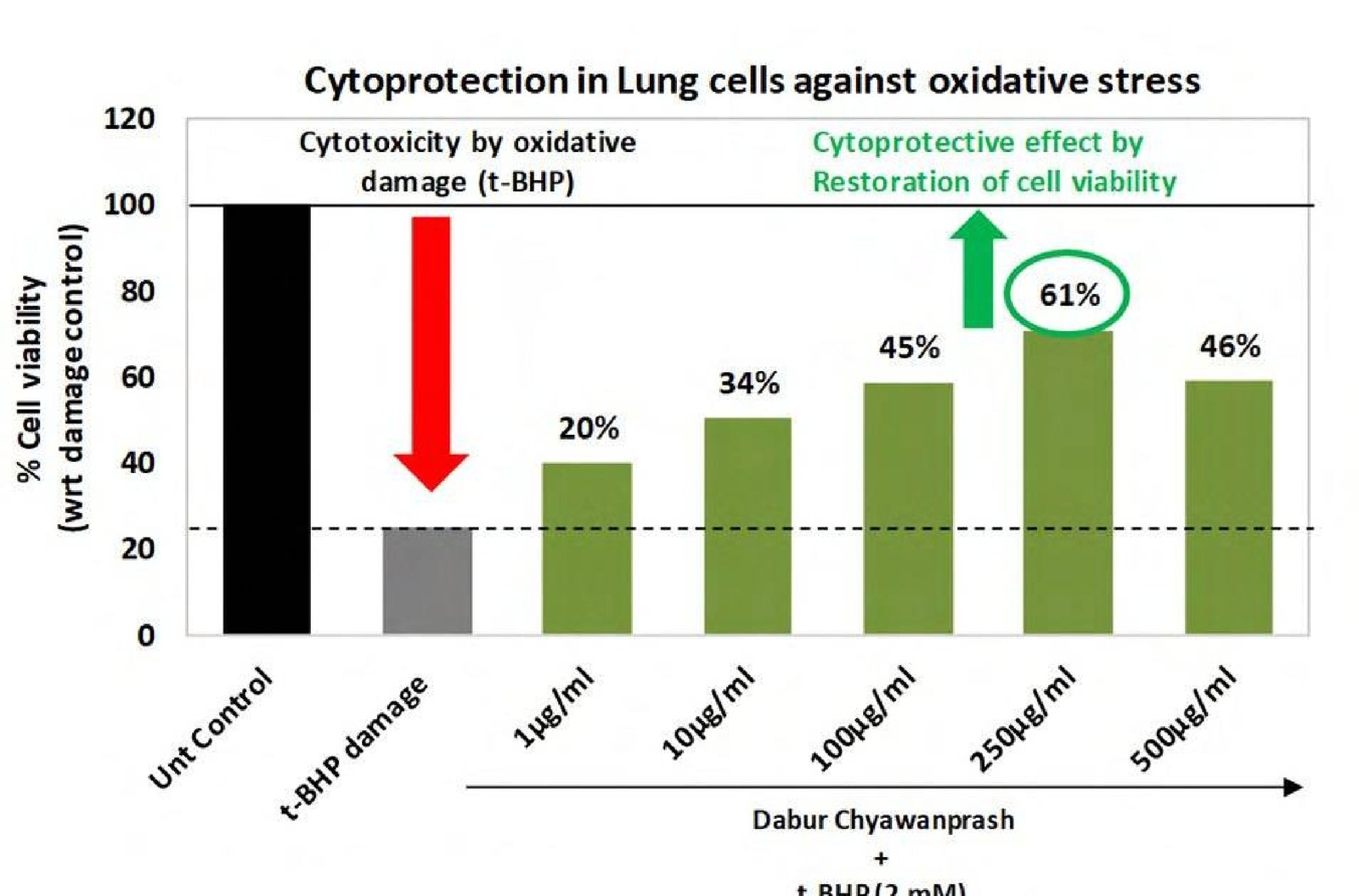 Study 1Cytoprotection in Lung CellsTo evaluate the effect of Dabur Chyawanprash (DCP) on respiratory health using in vitro cytoprotection assays.
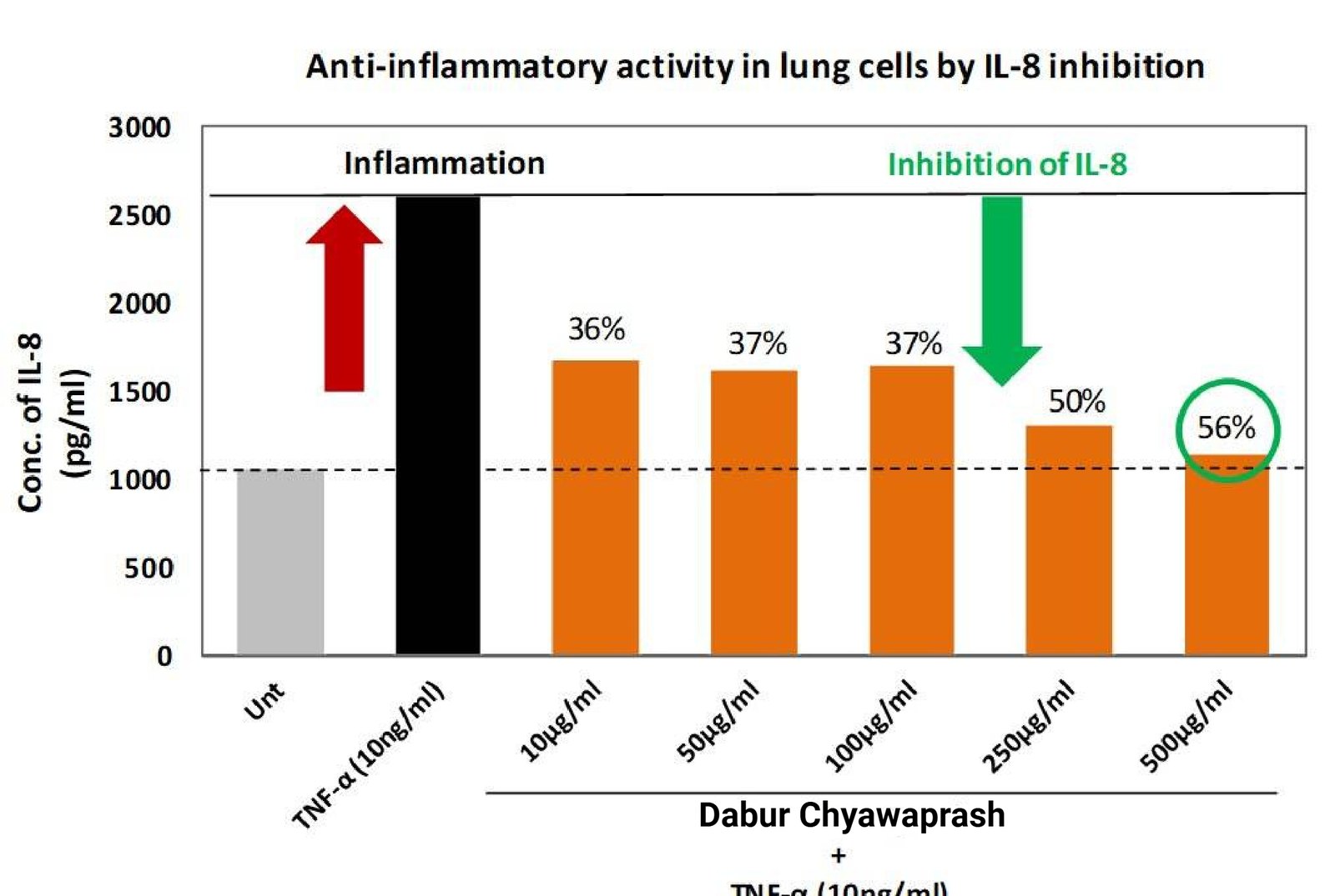 Study 2Anti-inflammatory Effect in Respiratory CellsTo evaluate the effect of Dabur Chyawanprash (DCP) on respiratory health using in vitro anti-inflammatory assays.
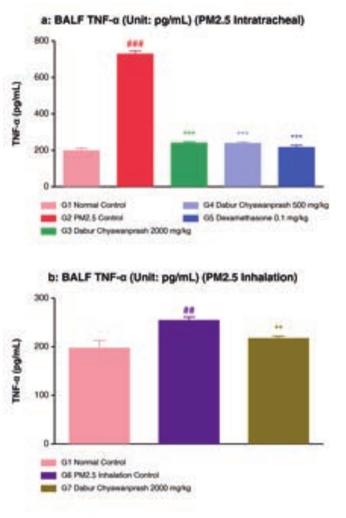 Study 3Anti-inflammatory and Immunomodulatory Activity of DCP on Particulate Matter-induced Pulmonary Disease in MiceTo evaluate the protective and prophylactic effect of Dabur Chyawanprash against particulate matter (PM2.5)-induced pulmonary disease in mice.
Mechanism
- 01Airborne irritants and particulate matter reach the epithelium
- 02Oxidative stress and inflammatory signalling
- 03DCP preserves viability and dampens IL-8, TNF-α and MMP-9
- 04Healthy airway surface and breathing comfort
Measured readouts
61%
At 250 µg/mL vs ~24% in t-BHP control
36–56%
TNF-α stimulated A549 cells, max at 500 µg/mL
~67%
66.9% and 67.1% at 2000 and 500 mg/kg
47.8–52.1%
PM2.5 intratracheal model (P < 0.001)
30.0–31.7%
PM2.5 intratracheal model (P < 0.01)
25.7%
PM2.5 inhalation model, 2000 mg/kg
Cellular benefits
- Supports respiratory health
- Helps protect respiratory cells from oxidative stress
- Helps maintain a balanced inflammatory response
- Helps maintain the integrity of lung cells
Interpretation
Cytoprotection and inflammatory moderation act together: protected epithelial cells reduce the stimulus for inflammation, and lower inflammation preserves the tissue.
Supports respiratory health and the natural defences of the airway.
DRF in-house respiratory cell assays and published PM2.5 mouse study.
Cytoprotection in Lung Cells
To evaluate the effect of Dabur Chyawanprash (DCP) on respiratory health using in vitro cytoprotection assays.
Human lung epithelial cells (A549). Cytoprotection – t-BHP induced oxidative damage
Methodology
Dabur Chyawanprash (DCP) was dissolved in DMSO to make stock of 200 mg/ml, which was further diluted in serum free medium to achieve final concentrations for treatment.
A total of 10,000 cells/well were seeded in a 96-well microplate and were incubated for 24 hours under standard cell-culture conditions to allow adequate cell attachment and stabilization.
On the following day, the cells were treated with Dabur Chyawanprash (DCP) at concentrations ranging from 1 to 500 μg/mL. The treatment was continued for an additional 24 hours to evaluate the potential cytoprotective effect of DCP against subsequent oxidative stress.
Following the 24-hour DCP treatment, the cells were exposed to tert-butyl hydroperoxide (tBHP) at a concentration of 2 mM for 3.5 hours. tBHP was used as an oxidative stress-inducing agent to generate cellular damage and establish an in vitro model of oxidative injury.
Appropriate untreated and tBHP-exposed control groups were included to assess basal cellular viability and the extent of oxidative-stress-induced cellular damage, respectively. The effect of DCP treatment was subsequently compared with the tBHP-induced damage control.
Following the oxidative stress exposure, cellular viability/cytoprotection was assessed using the MTT assay. The assay was based on the metabolic conversion of MTT into formazan by metabolically active cells, with the resulting signal being used as an indicator of cellular viability.
The absorbance generated by the treated and control groups was measured spectrophotometrically, and the relative cellular viability was determined. The extent of cytoprotection provided by DCP against tBHP-induced cellular damage was evaluated based on the increase in cellular viability relative to the tBHP-treated control.
The observed increase in cellular viability following DCP treatment, compared with cells exposed to tBHP alone, was interpreted as evidence of cytoprotective activity against chemically induced oxidative stress.
The percentage cell viability was calculated as = 100-%cytotoxicity. % Cytotoxicity was calculated as – ((Absorbance of untreated – Absorbance of treated)/(Absorbance of Untreated))*100. % Cytoprotection was calculated as = {(Absorbance of DCP-treated – Absorbance of t-BHP)/(Absorbance of untreated – Absorbance of t-BHP}*100
Restoration of cell viability as compared to t-BHP induced control, was considered indicative of cytoprotective response.
Key biomarkers
A t-BHP induced oxidative stress model was used to evaluate cellular resilience and cytoprotection against chemically induced oxidative damage. t-BHP exposure induced oxidative stress and reduced cellular viability, providing a suitable model for assessing the ability of test interventions to protect and maintain cellular integrity under oxidative stress conditions.
Results
Exposure to 2 mM tBHP markedly reduced cellular viability from 100% in untreated control cells to approximately 24%, confirming substantial oxidative stress-induced cellular damage.
Pretreatment with DCP resulted in partial restoration of cell viability across the tested concentrations. Cell viability increased to 20%, 34%, 45%, 61%, and 46% at 1, 10, 100, 250, and 500 μg/mL, respectively.
The maximum cytoprotective response was observed at 250 μg/mL, where viability reached 61%, indicating a substantial recovery from tBHP-induced damage. The response decreased at 500 μg/mL, suggesting that the protective effect did not increase further at the highest concentration tested. Overall, the findings demonstrated that DCP helped preserve cellular viability and attenuate oxidative stress-induced cellular damage, with the strongest effect observed at 250 μg/mL.
Scientific interpretation
These findings indicated that DCP attenuated tBHP-induced loss of cellular viability and supported cellular resilience under oxidative stress conditions. The restoration of viability following DCP pretreatment provided in vitro evidence of a cytoprotective effect against chemically induced oxidative damage, with the strongest response observed at 250 μg/mL under the tested conditions.
Scientific interpretation for consumer benefit claim
The cytoprotection study demonstrated that DCP attenuated the loss of cellular viability induced by oxidative stress. Following tBHP challenge, DCP treatment resulted in restoration of cellular viability, where viability reached 61% compared with approximately 24% in the tBHP-damaged control. These findings indicated that DCP helped maintain cellular integrity and resilience under oxidative stress conditions. Collectively, the results provided in vitro mechanistic support for a cytoprotective benefit, particularly in relation to protection of respiratory cells against oxidative stress.
Below consumer claims can be made:
- Helps protect respiratory cells from oxidative stress
- Supports respiratory cellular health
- Helps maintain healthy respiratory cells
- Supports cellular resilience for respiratory health
- Helps protect respiratory cells against oxidative damage t-BHP 2 mM
Anti-inflammatory Effect in Respiratory Cells
To evaluate the effect of Dabur Chyawanprash (DCP) on respiratory health using in vitro anti-inflammatory assays.
Human lung epithelial cells (A549). Anti-inflammatory – TNF-α induced inflammation.
Methodology
Dabur Chyawanprash (DCP) was dissolved in DMSO to make stock of 200 mg/ml, which was further diluted in serum free medium to achieve final concentrations for treatment.
A total of 0.1 million cells (1 × 10⁵ cells/well) were seeded in a 24-well tissue-culture plate and were incubated for 24 hours under standard cell-culture conditions to allow adequate cell attachment and stabilization.
Following the initial incubation, the cells were subjected to serum starvation for 24 hours. Serum starvation was performed to minimize the influence of serum-derived growth factors and basal inflammatory signaling and to establish a more controlled cellular response to the subsequent treatments.
After serum starvation, the cells were treated with Dabur Chyawanprash (DCP) at concentrations ranging from 10 to 500 μg/mL. The treatment was continued for 24 hours to assess the potential modulatory effect of DCP on the inflammatory response.
Following the 24-hour DCP treatment, inflammation was experimentally induced by addition of TNF-α at a concentration of 10 ng/mL. The cells were subsequently incubated for an additional 24 hours to allow TNF-α-mediated induction of the inflammatory response.
Appropriate experimental controls were maintained, including untreated cells and TNF-α-stimulated control cells, to establish basal and induced levels of inflammatory response, respectively. The DCP-treated, TNF-α-stimulated groups were compared with the TNF-α control group to determine the extent of modulation by DCP.
At the end of the TNF-α stimulation period, the cell-culture supernatants were carefully collected from the respective wells and were processed/stored under appropriate conditions until analysis.
The concentration of interleukin-8 (IL-8) in the collected cellculture supernatants was quantified using a specific ELISA, according to the manufacturer’s recommended procedure. IL-8 was selected as a key inflammatory mediator to assess the inflammatory response induced by TNF-α and its modulation following DCP treatment.
The IL-8 concentrations measured in the DCP-treated groups were compared with those observed in the TNF-α-stimulated control. The percentage inhibition of TNF-α-induced IL-8 production was calculated to determine the extent of anti-inflammatory activity exhibited by DCP.
% Inhibition of IL-8 = [(IL-8 concentration in TNF-α control − IL-8 concentration in DCP-treated group) / IL-8 concentration in TNF-α control] × 100
A reduction in IL-8 secretion following DCP treatment, compared with the TNF-α-stimulated control, was considered indicative of inhibition of the inflammatory response.
Key biomarkers
A TNF-α-induced inflammatory model was used to evaluate modulation of inflammatory signaling in respiratory cells. TNF-α stimulation induced the secretion of IL-8, an important pro-inflammatory mediator associated with respiratory inflammation. Changes in IL-8 secretion therefore provided a measure of the ability of test interventions to modulate the cellular inflammatory response.
Results
TNF-α stimulation markedly increased IL-8 secretion from approximately 1,000 pg/mL in untreated cells to around 2,600 pg/mL, confirming the induction of an inflammatory response.
Treatment with DCP resulted in a reduction in TNF-α-induced IL-8 production across the tested concentration range of 10–500 μg/mL. At 10 μg/mL, DCP reduced IL-8 levels to approximately 1,670 pg/mL, corresponding to 36% inhibition. IL-8 inhibition remained consistent at 50 and 100 μg/mL, with approximately 37% inhibition observed at both concentrations.
A further reduction in IL-8 production was observed at 250 μg/mL, where inhibition increased to 50%. The maximum response was observed at 500 μg/mL, where IL-8 levels were reduced to approximately 1,150 pg/mL, corresponding to 56% inhibition compared with the TNF-α control.
Overall, DCP demonstrated a progressive reduction in TNF-α-induced IL-8 secretion, particularly at the higher concentrations, indicating modulation of the inflammatory response in the lung-cell model.
Scientific interpretation
The results demonstrated that DCP modulated the inflammatory response induced by TNF-α in lung cells, as evidenced by the reduction in IL-8 secretion. TNF-α stimulation markedly elevated IL-8 levels, while DCP treatment reduced IL-8 production by 36–56% across the tested concentration range. The inhibitory effect became more pronounced at higher concentrations, reaching a maximum of 56% inhibition at 500 μg/mL. Since IL-8 is an important mediator of inflammatory signaling and is involved in the recruitment and activation of inflammatory cells, its reduction indicated attenuation of TNF-α-driven inflammatory signaling. Collectively, the findings provided in vitro mechanistic evidence for the anti-inflammatory potential of DCP and its ability to support maintenance of a balanced inflammatory response relevant to respiratory cellular health.
Scientific interpretation for consumer benefit claim
The reduction in TNF-α-induced IL-8 secretion by 36–56% demonstrated that DCP modulated an important inflammatory signaling pathway in lung cells. The maximum 56% inhibition of IL-8 at 500 μg/mL indicated attenuation of the inflammatory response under the experimental conditions. As IL-8 is an important mediator of inflammatory signaling in respiratory tissues, these findings provided in vitro mechanistic support for maintaining a balanced inflammatory response and supporting respiratory cellular health.
Below consumer claims can be made:
- Supports a healthy inflammatory response.
- Helps maintain a balanced inflammatory response.
- Supports respiratory health by helping maintain a healthy inflammatory response.
- Helps support healthy respiratory function.
- Supports the body’s natural anti-inflammatory defence.
- Helps maintain respiratory cellular health.
Anti-inflammatory and Immunomodulatory Activity of DCP on Particulate Matter-induced Pulmonary Disease in Mice
To evaluate the protective and prophylactic effect of Dabur Chyawanprash against particulate matter (PM2.5)-induced pulmonary disease in mice.
PM2.5 exposure-induced pulmonary disease in the Balb/c mouse, by intratracheal instillation and by repeated aerosol inhalation.
Method
56 male Balb/c mice were divided into 7 groups (n = 8 per group). G1 served as normal control and G2 as PM2.5 disease control. G3 and G4 received Dabur Chyawanprash at 2000 and 500 mg/kg respectively, G5 received dexamethasone 0.1 mg/kg, and G6 served as PM2.5 inhalation control. G7 received DCP 2000 mg/kg with PM2.5 inhalation.
DCP was administered orally at 500 and 2000 mg/kg once daily for 38 days. The study was designed as a prophylactic model, with treatment beginning before PM2.5 exposure. Dexamethasone 0.1 mg/kg was used as the standard drug.
After completion of the treatment duration, blood was collected for hematology and serum IgE. Animals were sacrificed humanely; bronchoalveolar lavage fluid (BALF) was collected for inflammatory cell counts and cytokine estimation, and lungs were collected for biochemical analysis and histopathology.
Study at a glance
SPECIES Male Balb/c mice, 56 animals
DOSE DCP 500 and 2000 mg/kg, oral, once daily
STANDARD DRUG Dexamethasone 0.1 mg/kg
SAMPLES Serum, BALF, lung homogenate, lung histology
Pm2.5 exposure models
Intratracheal: 100 mg PM2.5 per mouse in 100 µL per animal, concentration 1 mg/mL in normal saline, administered on Days 29 and 35.
Inhalation: PM2.5 aerosol at 2 mg / 5 mL for 15 minutes in a histamine chamber on Days 29, 31, 33, 35 and 37.
Key parameters
Serum: IgE.
BALF (ELISA): TNF-α, IFN-γ, IL-17A, IL-6, and differential inflammatory cell counts (neutrophils, lymphocytes, eosinophils and other inflammatory cells).
Lung homogenate (PBS): MMP-9, histamine, IL-6 and TNF-α.
Histopathology: Lung fixed in 10% neutral buffered formalin, paraffin-embedded, H&E stained.
Other Parameters: Body-weight change, hematology, lung histopathological score, alveolar inflammatory infiltration, atelectasis, foamy histiocytes, perivascular fibrosis, alveolar edema, peribronchial lymphoid hyperplasia.
GROUPS 7 groups, n = 8 per group
DURATION 38 days, prophylactic design
EXPOSURE PM2.5 intratracheal (Days 29, 35) and aerosol inhalation (Days 29–37)
SAFETY READOUTS Body weight, hematology, clinical signs
Results
Inflammatory and immunomarkers in BALF
Dabur Chyawanprash demonstrated protective effects against PM2.5-induced pulmonary inflammation in mice. The PM2.5 disease-control group showed increased inflammatory markers and pulmonary changes compared with the normal control. DCP treatment at 500 and 2000 mg/kg reduced several of these PM2.5-induced changes.
BALF TNF-α DCP 2000 and 500 mg/kg reduced TNF-α by 66.9% and 67.1% respectively versus disease control (p < 0.001). In the inhalation model, DCP 2000 mg/kg produced a significant 14.7% reduction versus the PM2.5 inhalation control.
Balf IL-17 and IL-6
In the inhalation model, DCP 2000 mg/kg significantly reduced BALF IL-17 by 25.7% (p < 0.05). BALF IL-6 showed a nonsignificant reduction with DCP at both doses in the intratracheal model. For the intratracheal model, DCP produced non-significant decreases in IFN-γ and IL-17 at the tested doses.
Differential cell count in BALF
In the intratracheal model, PM2.5 increased BALF neutrophils, while DCP produced reductions in neutrophil infiltration; some changes were not statistically significant. Lymphocytes were significantly decreased by PM2.5; DCP produced non-significant increases in lymphocytes level. Eosinophil changes were not consistently significant. In the inhalation model, DCP 2000 mg/kg produced a non-significant reduction in neutrophils compared with the PM2.5 inhalation control.
Immunomarker in serum
Serum IgE: DCP 2000 and 500 mg/kg produced 27.3% and 14.7% reductions respectively, although these changes were not statistically significant.
Inflammatory and immunomarkers in lung tissue
LUNG TNF-α
DCP 2000 and 500 mg/kg reduced lung TNF-α by 22.7% and 26.2% respectively (p < 0.01). In the inhalation model, DCP 2000 mg/kg produced a 34.1% reduction (p < 0.001).
Lung MMP-9
DCP 2000 and 500 mg/kg reduced MMP-9 by 47.8% and 52.1% respectively (p < 0.001). DCP 2000 mg/kg also reduced MMP-9 by 46.1% in the inhalation model (p < 0.001).
Lung histamine and IL-6
In the intratracheal model, PM2.5 significantly increased lung histamine by 38.5%. DCP reduced lung histamine by 31.7% at 2000 mg/kg (p < 0.01) and 30.0% at 500 mg/kg (p < 0.01). PM2.5 also significantly increased lung IL-6 by 77.0%; DCP produced nonsignificant reductions in the intratracheal model, while in the inhalation model DCP 2000 mg/kg significantly reduced lung IL-6 by 30.9% (p < 0.01).
Histopathology
DCP 500 mg/kg produced a significant reduction in the total histological score compared with the PM2.5 disease-control group (p < 0.05); DCP 2000 mg/kg showed a non-significant decrease. For alveolar neutrophil infiltration, DCP 500 mg/kg produced a significant reduction (p < 0.05) versus PM2.5 disease control. Other findings included atelectasis, foamy histiocytes, perivascular fibrosis, alveolar edema and peribronchial lymphoid hyperplasia.
Body weight and tolerability
PM2.5 disease-control animals showed a 12.6% body-weight loss during Days 36–38, whereas DCP-treated animals did not show significant body-weight reduction. No clinical signs of toxicity were observed in DCP-treated groups.
Conclusion
The study demonstrated that PM2.5 exposure produced pulmonary inflammatory changes, including increases in inflammatory cytokines and lung inflammatory markers, alterations in BALF inflammatory cells, and histopathological changes.
Prophylactic administration of Dabur Chyawanprash, particularly at 500 and 2000 mg/kg, attenuated several of these PM2.5-associated changes. The effects were most consistently demonstrated for TNF-α, MMP-9, selected cytokines, inflammatory cell infiltration and histopathology.
The study concluded that Dabur Chyawanprash demonstrated a prophylactic protective effect against PM2.5 - induced pulmonary pathology in the experimental mouse model. The reported benefit was associated with inhibition of inflammatory cytokines, lung MMP-9, inflammatory cell infiltration and histopathological changes.
Scientific interpretation
The study demonstrates that Dabur Chyawanprash has prophylactic anti-inflammatory and immunomodulatory activity against PM2.5 - induced pulmonary inflammation in mice. Administration at 500 and 2000 mg/kg attenuated several PM2.5 - associated changes, particularly reductions in BALF and lung TNF-α, lung MMP-9, selected inflammatory cytokines, inflammatory-cell infiltration, and histopathological changes.
The effects indicate that DCP may help modulate inflammatory responses and protect pulmonary tissue from PM2.5-induced injury in this experimental model. The study provides evidence of reduced inflammatory markers and pulmonary pathological changes following DCP administration.
Scientific interpretation for consumer benefit claim
Helps protect and support lung health from environmental particulate stress.
Helps maintain healthy lungs and a balanced inflammatory response.
Source
Kumar S, Rugvedi P, Mani K, Gupta A. Evaluation of anti-inflammatory matter induced pulmonary disease in mice. Journal of Ayurveda and and immunomodulatory activity of Chyawanprash on particulate Integrative Medicine. 2021;12:649–656
Shielding liver cells against oxidative insult
Substantiated by Oxidative Damage Protection Evidence in Hepatocytes For Energy & Stamina
Hepatocytes carry the metabolic and detoxification load and are correspondingly exposed to oxidative injury. DCP preserves their viability under t-BHP challenge.
Model systemHepG2 human liver cells under 250 µM t-BHP
1 study in this area
Mechanism
- 01Hepatotoxic and oxidative challenge
- 02Membrane and mitochondrial stress in hepatocytes
- 03DCP cytoprotection and antioxidant support
- 04Preserved hepatocyte function
Measured readouts
~47 → ~86%
Restored at 250–500 µg/mL
~74%
Plateau reached at 250–500 µg/mL
~44%
Cytoprotection, viability ~71%
~34%
Cytoprotection, viability ~65%
~10%
Cytoprotection, viability ~52%
~47%
Viability in the oxidative stress control
Cellular benefits
- Supports liver health
- Protects liver cells from oxidative stress
- Supports cellular protection against oxidative damage
- Helps maintain cellular health and resilience
Interpretation
The response is clearly concentration dependent and plateaus at 250 µg/mL, the profile expected of a genuine protective effect rather than assay noise.
Supports liver health and protects liver cells from oxidative stress.
DRF in-house hepatocyte assays.
Cytoprotective effect on liver
To evaluate the cytoprotective effect of Dabur Chyawanprash (DCP) on liver health using in vitro assays.
Human liver cells (HepG2). Cytoprotection – t-BHP induced oxidative damage
Methodology
Dabur Chyawanprash (DCP) was dissolved in DMSO to make stock of 200 mg/ml, which was further diluted in serum free medium to achieve final concentrations for treatment.
The cytoprotective potential of Dabur Chyawanprash (DCP) was evaluated using an in vitro oxidative stress model in liver cells. Briefly, 10,000 cells were seeded per well in a 96-well microplate and allowed to adhere and stabilize for 24 hours under standard cell culture conditions.
Following cell attachment, the cells were treated with DCP at concentrations ranging from 0.1–100 μg/mL for 24 hours to assess its potential protective effect. After the treatment period, oxidative stress was induced by exposing the cells to 250 μM tertbutyl hydroperoxide (t-BHP) for 3.5 hours. t-BHP is a commonly used chemical inducer of oxidative stress that promotes intracellular oxidative damage and consequently reduces cellular viability.
Following oxidative stress induction, cell viability and cytotoxicity were quantified using the MTT assay, which measures the metabolic activity of viable cells based on their ability to reduce MTT to insoluble formazan. The absorbance of the resulting formazan product was measured and used as an indicator of cellular metabolic viability.
The cytoprotective effect of DCP was evaluated by comparing the viability of DCP-pretreated, t-BHP-challenged cells with that of the t-BHP-exposed control cells. An increase in cell viability in DCP-treated cells relative to the oxidative stress control was interpreted as evidence of protection against t-BHP-induced cellular damage.
The percentage cytoprotection was calculated with respect to the t-BHP-exposed control, allowing the protective effect of DCP against chemically induced oxidative stress to be quantitatively assessed. Collectively, this model provides an in vitro assessment of the ability of DCP to preserve liver-cell viability and enhance cellular resilience under oxidative stress conditions.
The percentage cell viability was calculated as = 100-%cytotoxicity. % Cytotoxicity was calculated as – ((Absorbance of untreated – Absorbance of treated)/(Absorbance of Untreated))*100. % Cytoprotection was calculated as = {(Absorbance of DCP-treated – Absorbance of t-BHP)/(Absorbance of untreated – Absorbance of t-BHP}*100
Restoration of cell viability as compared to t-BHP induced control, was considered indicative of cytoprotective response.
Key biomarkers
A tBHP-induced oxidative stress model was used to evaluate cellular resilience and cytoprotection against chemically induced oxidative damage. tBHP exposure induced oxidative stress and reduced cellular viability, providing a suitable model for assessing the ability of test interventions to protect and maintain cellular integrity under oxidative stress conditions.
Results
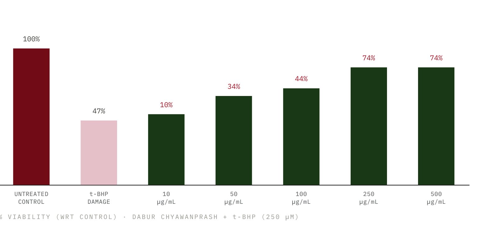
The t-BHP challenge produced a marked reduction in HepG2 cell viability, with viability decreasing from 100% in untreated control cells to approximately 47% following exposure to 250 μM t-BHP, confirming successful induction of oxidative cytotoxicity.
Pretreatment with Dabur Chyawanprash (DCP) demonstrated a concentration-dependent restoration of cell viability following oxidative stress:
At 10 μg/mL DCP, cell viability increased to approximately 52%, corresponding to ~10% cytoprotection relative to the t-BHP control.
At 50 μg/mL, viability increased further to 65%, representing ~34% cytoprotection.
At 100 μg/mL, viability reached approximately 71%, corresponding to ~44% cytoprotection.
At 250 μg/mL, viability increased substantially to approximately 86%, corresponding to ~74% cytoprotection.
Increasing DCP to 500 μg/mL maintained viability at approximately 86%, with cytoprotection remaining at ~74%, suggesting that the protective effect had reached a plateau.
Scientific interpretation
Overall, DCP demonstrated a clear concentration-dependent cytoprotective response against t-BHP-induced oxidative damage in HepG2 cells. The progressive recovery of cell viability from ~47% in the oxidative stress control to ~86% following DCP treatment indicates that DCP substantially attenuated oxidative-stress-associated cellular injury.
Scientific interpretation for consumer benefit claim
The study demonstrates that Dabur Chyawanprash (DCP) protects liver cells against oxidative stress-induced damage. t-BHP exposure substantially reduced HepG2 cell viability, while DCP treatment progressively restored cell viability, with up to ~74% cytoprotection observed at 250–500 μg/mL.
These findings indicate that DCP may help enhance cellular resilience against oxidative stress and protect cells from oxidative damage.
Cytoprotection in HepG2 cells against oxidative
Potential consumer benefit claims:
- Helps protect cells against oxidative stress
- Supports cellular protection against oxidative damage
- Helps maintain cellular health and resilience
- Provides cellular protection against oxidative stress
- Supports liver-cell protection against oxidative damage
Damage
Raising cellular energy availability
Substantiated by ATP Level Enhancement in PBMCs – Evidence for Cellular Energy and Vitality
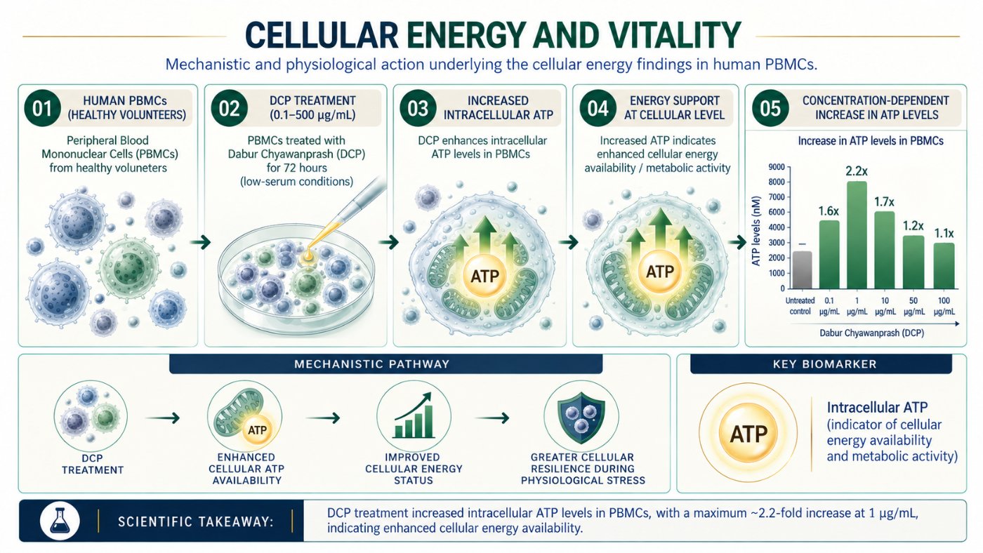
ATP is the energy currency of every cell. In blood immune cells from healthy volunteers, DCP raised intracellular ATP after 72 hours in low-serum culture.
Model systemHuman PBMCs from healthy volunteers, 72 h, read against untreated control
1 study in this area
Mechanism
- 01DCP treatment of immune cells
- 02Enhanced cellular ATP availability
- 03Improved cellular energy status
- 04Greater resilience during physiological stress
Measured readouts
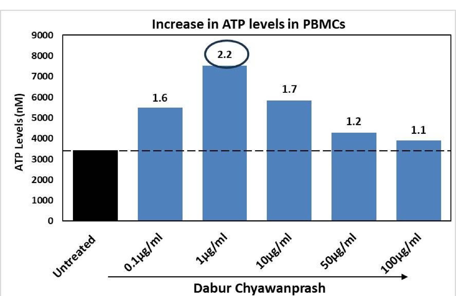
2.2×
Maximum response vs untreated cells
1.7×
Intracellular ATP
1.6×
Intracellular ATP
1.2×
Intracellular ATP
1.1×
Intracellular ATP
0.1–500 µg/mL
72 h, 0.1% FBS, luminescence ATP assay
Cellular benefits
- Supports cellular energy and vitality
- Supports natural energy metabolism
- Helps maintain cellular energy levels
- Supports cellular resilience during physiological stress
Interpretation
Higher ATP indicates more energy available for immune-cell activation and maintenance. On its own it is not evidence of improved stamina or reduced fatigue.
Supports cellular energy and vitality.
DRF in-house PBMC ATP assay.
Energy boost potential using PBMCs
To evaluate the energy boost potential of Dabur Chyawanprash (DCP) by ATP assay in human Peripheral Blood Mononuclear Cells (PBMCs).
Human Peripheral Blood Mononuclear Cells (PBMCs) from healthy volunteers
Methodology
Dabur Chyawanprash (DCP) was dissolved in DMSO to make stock of 200 mg/ml, which was further diluted in serum free medium to achieve final concentrations for treatment.
0.2 million PBMCs were seeded in each well of a 24-well plate in RPMI medium containing 0.1% FBS and allowed to stabilize for 2 hours.
The PBMCs were then treated with Dabur Chyawanprash (DCP) at 0.1–500 μg/mL for 72 hours under low-serum conditions to evaluate its effect on cellular energy status.
Following treatment, the cells were harvested and cell lysates were prepared for quantification of intracellular ATP.
ATP levels were measured using a validated ATP assay kit, with intracellular ATP serving as an indicator of cellular energy availability and metabolic activity.
The ATP levels in DCP-treated PBMCs were compared with untreated control cells, and the percentage increase in intracellular ATP was determined.
An increase in ATP following DCP treatment indicates enhanced cellular energy availability/metabolic activity, suggesting that DCP may support the energy requirements of metabolically active immune cells.
From a biological perspective, adequate ATP availability is essential for immune-cell activation, proliferation, cytokine production, cellular maintenance and other energy-demanding cellular processes.
Therefore, increased ATP levels in PBMCs provide mechanistic evidence for a potential cellular energy-supporting effect of DCP. However, this assay demonstrates increased cellular ATP and should not by itself be interpreted as direct evidence of improved physical stamina or reduced fatigue.
Fold increase in ATP levels was calculated as = ((Luminescence of DCP treated cells – Luminescence of untreated cells)/(Luminescence of Untreated cells))*100
Key biomarkers
This assay is biologically relevant because ATP is the primary cellular energy currency required for virtually all energy-dependent processes. Measuring intracellular ATP in PBMCs provides an indication of their metabolic and energy status. Increased ATP following DCP treatment may indicate improved cellular energy availability and metabolic resilience, which can support energydemanding functions of immune cells such as activation, proliferation, cytokine production and cellular maintenance. Since immune responses and cellular repair processes require substantial energy, maintaining adequate ATP may contribute to overall cellular resilience during physiological stress. Thus, the assay provides mechanistic evidence for a potential energysupporting and cellular vitality benefit of DCP.
Increased intracellular ATP following DCP treatment indicates that DCP can support cellular energy availability and metabolic activity. ATP is required for energy-demanding processes including cellular maintenance, biosynthesis, membrane transport and immune-cell activation. Maintaining adequate ATP availability may help cells cope with metabolic and physiological stress and sustain normal cellular functions.
Mechanistic pathway
DCP enhanced cellular ATP availability improved cellular energy status greater cellular resilience during physiological stress potential support for vitality and stamina
Results
DCP treatment resulted in an increase in intracellular ATP levels in PBMCs compared with the untreated control, indicating an enhancement of cellular energy status. The strongest response was observed at 1 μg/mL, where ATP levels increased approximately 2.2-fold compared with untreated cells. ATP levels were also elevated at 0.1 μg/mL (1.6-fold), 10 μg/mL (1.7-fold), 50 μg/mL (1.2-fold), and 100 μg/mL (1.1-fold).
Overall, DCP demonstrated a positive effect on cellular ATP availability across the tested concentration range, with the maximum response at 1 μg/mL. Since ATP is the primary energy currency required for cellular maintenance, activation, biosynthesis and other energy-demanding processes, these findings suggest that DCP may support cellular energy metabolism and metabolic resilience.
From an energy and vitality perspective, the data provide mechanistic support for the potential of DCP to support cellular energy availability, which may contribute to maintaining cellular function during physiological stress.
Scientific interpretation
The results demonstrate that DCP enhances intracellular ATP availability in PBMCs, with the maximum response of approximately 2.2-fold at 1 μg/mL compared with untreated cells. ATP is the primary cellular energy currency and is essential for maintaining cellular homeostasis, membrane integrity, biosynthetic processes and energy-demanding immune-cell functions.
Scientific interpretation for consumer benefit claim
The increased intracellular ATP levels in PBMCs provide mechanistic evidence that DCP may support cellular energy availability and metabolic activity. The maximum 2.2-fold increase in ATP suggests enhanced energy status, which is important for maintaining cellular functions, immune-cell activity and resilience during physiological demands.
From a consumer-health perspective, these findings support a potential role of DCP in supporting cellular energy, vitality and resilience, providing a biological basis for an energy-support narrative. When combined with additional evidence related to metabolism, physical performance or fatigue, this mechanism could contribute to substantiation of broader energy and stamina benefits.
Potential consumer benefit claims:
- Supports cellular energy and vitality
- Supports natural energy metabolism
- Helps maintain cellular energy levels
- Supports cellular resilience during physiological stress
- Helps support energy availability for normal cellular functions
- Supports overall vitality and metabolic health
- Key scientific message: DCP cellular ATP support for energy, vitality & stamina.
Protecting muscle cells and their energy supply
Substantiated by Oxidative Stress & Hypoxia Protection Evidence in Muscle Cells
Two mechanisms were examined in muscle cells: direct cytoprotection against oxidative and hypoxic damage, and preservation of mitochondrial membrane potential.
Model systemC2C12 myoblasts under t-BHP (75 µM) and CoCl₂ (420.3 µM), 48 h
2 studies in this area
 Study 1Cytoprotective effect in muscle cellsTo evaluate the muscle strengthening potential of Dabur Chyawanprash (DCP) in muscle cells by cytoprotective effect.
 Study 2Restoration of Mitochondrial Membrane Potential Against StressTo evaluate the muscle strengthening potential of Dabur Chyawanprash (DCP) in muscle cells by restoration of mitochondrial membrane potential against stress.
Mechanism
- 01Oxidative and hypoxic damage to muscle cells
- 02Myocyte stress and mitochondrial depolarisation
- 03DCP cytoprotection and mitochondrial support
- 04Muscle-cell integrity preserved
Measured readouts
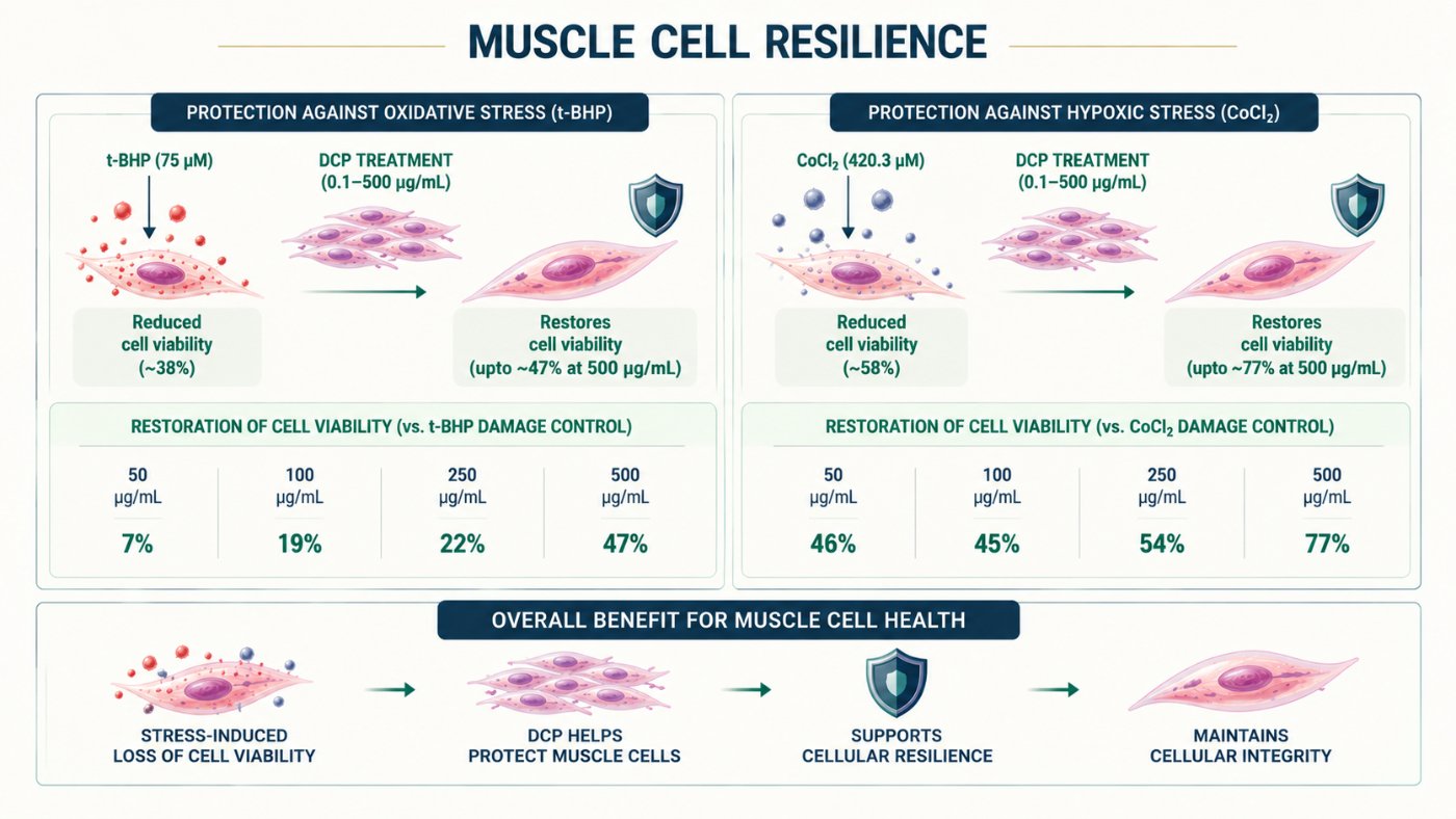
47%
Restoration at 500 µg/mL (damage control ~38%)
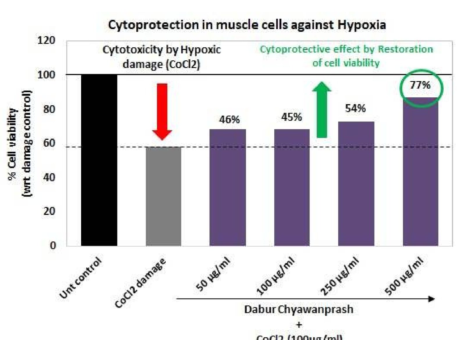
77%
Restoration at 500 µg/mL (damage control ~58%)
+202%
Membrane potential at 500 µg/mL, JC-1
+118%
Membrane potential at 500 µg/mL, JC-1
7 → 47%
50, 100, 250, 500 µg/mL: 7, 19, 22, 47%
46 → 77%
50, 100, 250, 500 µg/mL: 46, 45, 54, 77%
Cellular benefits
- Helps protect muscle cells from cellular stress
- Supports muscle-cell resilience against oxidative stress
- Helps maintain mitochondrial integrity under stress
- Supports cellular resilience and vitality
Interpretation
Cytoprotection and energetic support are complementary: preserved mitochondria supply the ATP that contractile function and repair both depend on.
Supports muscle strengthening potential by protecting muscle cells from cellular stress.
DRF in-house muscle cell assays.
Cytoprotective effect in muscle cells
To evaluate the muscle strengthening potential of Dabur Chyawanprash (DCP) in muscle cells by cytoprotective effect.
Mouse Myoblast cells (C2C12). Cytoprotection – t-BHP induced oxidative damage; CoCl₂ induced hypoxic stress
Methodology
Dabur Chyawanprash (DCP) was dissolved in DMSO to make stock of 200 mg/ml, which was further diluted in serum free medium to achieve final concentrations for treatment.
0.01 million cells (1 × 10⁴ cells/well) were seeded in 96-well plates containing culture medium supplemented with 10% FBS. The cells were incubated for 24 h under standard cell-culture conditions to allow proper cell attachment and stabilization.
Following the 24-h incubation, cells were exposed to t-BHP (75 μM) or CoCl₂ (420.3 μM) to induce cellular stress and reduce cell viability, thereby establishing the respective damage-control models.
Cells were simultaneously treated with Dabur Chyawanprash (DCP) at concentrations ranging from 0.1–500 μg/mL in the presence of either t-BHP or CoCl₂. The treatment was continued for 48 h.
Appropriate controls were maintained, including an untreated control representing normal cell viability and a t-BHP/CoCl₂ damage control representing stress-induced loss of viability in the absence of DCP.
After 48 h of treatment, cellular viability was assessed using the MTT assay. The metabolically active viable cells reduce MTT to purple-colored formazan crystals, and the intensity of the developed color corresponds to cellular metabolic activity and viability.
The absorbance of the formazan product was measured using a microplate reader. The viability of cells treated with DCP in combination with t-BHP or CoCl₂ was compared with the respective damage-control group.
The cytoprotective effect of DCP was determined based on the restoration of cell viability relative to the corresponding t-BHP/CoCl₂-induced damage control. Higher restoration of viability indicates greater protection against stress-induced cellular injury.
Overall, the assay evaluates the ability of DCP to attenuate stress-induced loss of muscle-cell viability and promote cellular resilience under t-BHP- and CoCl₂-induced injury conditions.
The percentage cell viability was calculated as = 100-%cytotoxicity. % Cytotoxicity was calculated as – ((Absorbance of untreated – Absorbance of treated)/(Absorbance of Untreated))*100. % Cytoprotection was calculated as = {(Absorbance of DCP-treated – Absorbance of t-BHP)/(Absorbance of untreated – Absorbance of t-BHP}*100
Restoration of cell viability as compared to t-BHP induced control, was considered indicative of cytoprotective response.
Key biomarkers
Cytoprotection – t-BHP-induced oxidative damage
A t-BHP-induced oxidative stress model was used to assess cellular resilience and cytoprotection against chemically induced oxidative damage. t-BHP exposure induces oxidative stress and reduces cellular viability, providing a suitable model for evaluating the ability of test interventions to protect cells and maintain cellular integrity under oxidative stress conditions.
Cytoprotection – CoCl₂ -induced hypoxic stress
A CoCl₂ -induced hypoxia model was used to evaluate cellular resilience and cytoprotection under chemically induced hypoxic stress. CoCl₂ exposure mimics hypoxia-like conditions, resulting in cellular stress and reduced viability, thereby providing a suitable model for assessing the ability of test interventions to preserve cell viability and cellular integrity under hypoxic conditions.
Results
In the t-BHP-induced oxidative stress model, exposure to 75 μM t-BHP markedly reduced muscle-cell viability to approximately 38% of the untreated control, confirming effective induction of oxidative cellular damage.
Co-treatment with Dabur Chyawanprash (DCP) demonstrated a progressive restoration of cell viability with increasing concentrations. At 50 μg/mL, DCP produced a 7% cytoprotective effect, which increased to 19% at 100 μg/mL and 22% at 250 μg/mL.
At the highest concentration tested (500 μg/mL), DCP showed the strongest protective response, with approximately 47% restoration of cell viability relative to the t-BHP-induced damage control. This indicates that DCP substantially attenuated the loss of viability caused by oxidative stress.
In the CoCl₂-induced hypoxic stress model, CoCl₂ treatment reduced muscle-cell viability to approximately 58% of the untreated control, demonstrating successful induction of hypoxia-associated cellular stress.
DCP treatment produced a marked restoration of cell viability in the CoCl₂-damaged cells. A 46% cytoprotective effect was observed at 50 μg/mL, with comparable protection of 45% at 100 μg/mL, followed by an increase to 54% at 250 μg/mL.
The maximum protective response was observed at 500 μg/mL DCP, where cytoprotection reached approximately 77%, indicating substantial restoration of cell viability toward the untreated control level.
Overall, DCP demonstrated cytoprotective activity in muscle cells under both oxidative stress (t-BHP) and hypoxia-mimicking stress (CoCl₂). The effect was particularly pronounced in the CoCl₂ model, with up to 77% restoration of viability, compared with 47% restoration under t-BHP-induced oxidative stress.
These findings suggest that DCP can attenuate stress-induced loss of muscle-cell viability and enhance cellular resilience, with the strongest protection observed at 500 μg/mL in both experimental models.
Scientific interpretation
DCP demonstrated significant cytoprotective activity in muscle cells exposed to oxidative (t-BHP) and hypoxiamimicking (CoCl₂) stress. DCP restored cell viability in a concentration-dependent manner, with maximum protection of 47% against t-BHP-induced damage and 77% against CoCl₂ -induced damage at 500 μg/mL. These findings indicate that DCP may enhance cellular resilience and protect muscle cells from stress-induced loss of viability.
Scientific interpretation for consumer benefit claim
DCP demonstrated in vitro cytoprotective activity in muscle cells, helping preserve cell viability under oxidative and hypoxia-like stress conditions. The restoration of viability observed with DCP suggests potential to support cellular resilience and help protect muscle cells against stress-induced damage, providing a scientific basis for a consumer benefit related to muscle-cell protection and resilience against cellular stress.
Potential consumer benefit claims:
- Helps protect muscle cells from cellular stress
- Supports muscle-cell resilience against oxidative stress
- Helps maintain muscle-cell health under stress
- Supports cellular protection against oxidative and hypoxia-like stress
- Helps protect cells against stress-induced damage
- Supports cellular resilience and vitality
Restoration of Mitochondrial Membrane Potential Against Stress
To evaluate the muscle strengthening potential of Dabur Chyawanprash (DCP) in muscle cells by restoration of mitochondrial membrane potential against stress.
Mouse Myoblast cells (C2C12)
Methodology
Dabur Chyawanprash (DCP) was dissolved in DMSO to make stock of 200 mg/ml, which was further diluted in serum free medium to achieve final concentrations for treatment.
0.01 million cells (1 × 10⁴ cells/well) were seeded in 96-well plates containing culture medium supplemented with 10% FBS and incubated for 24 h to allow cell attachment and stabilization.
Following the initial incubation, cells were subjected to cellular stress using t-BHP (75 μM) or CoCl₂ (420.3 μM). These treatments were used to induce oxidative and hypoxia-mimicking stress, respectively, resulting in disruption of normal mitochondrial function.
Cells were co-treated with DCP at concentrations ranging from 0.1–500 μg/mL in the presence of either t-BHP or CoCl₂ and incubated for a further 48 h. Appropriate untreated and stress-induced damage controls were included.
After 48 h, mitochondrial membrane potential (ΔΨm) was assessed using the JC-1 fluorescent dye assay. JC-1 accumulates within healthy mitochondria and forms aggregates that exhibit red fluorescence, whereas loss of mitochondrial membrane potential results predominantly in JC-1 remaining in its monomeric form, producing green fluorescence.
Fluorescence intensity was measured using appropriate excitation/emission settings: 528/590 nm for red fluorescence and 485/528 nm for green fluorescence. The red-to-green fluorescence ratio was used as an indicator of mitochondrial membrane potential, with a higher ratio reflecting better preservation of mitochondrial polarization.
The mitochondrial membrane potential in DCP-treated cells was compared with the respective t-BHP/CoCl₂ damage-control group. The percentage increase in mitochondrial membrane potential was calculated relative to the corresponding stress-induced damage control.
An increase in the red/green fluorescence ratio following DCP treatment indicates restoration or preservation of mitochondrial membrane potential, suggesting that DCP may help protect mitochondrial function against stress-induced mitochondrial depolarization.
Mitochondrial Membrane potential (MMP) was calculated as ratio of Red:Green ratio. % inhibition in MMP was calculated as – ((Red:Green ratio of untreated – Red:Green ratio of treated)/(Red:Green ratio of Untreated))*100
Key biomarkers
The JC-1 assay provides a mechanistic measure of mitochondrial health by assessing changes in mitochondrial membrane potential under cellular stress. Preservation of the red/green fluorescence ratio following DCP treatment indicates maintenance of mitochondrial integrity and function, supporting DCP’s potential to promote cellular energy homeostasis, muscle-cell resilience, and protection against stress-induced mitochondrial dysfunction.
Results
In the t-BHP-induced oxidative stress model, treatment with 75 μM t-BHP caused a pronounced reduction in mitochondrial membrane potential, indicating substantial mitochondrial depolarization and disruption of mitochondrial function.
DCP treatment progressively restored mitochondrial membrane potential. At 50 μg/mL and 100 μg/mL, DCP produced 12% and 23% increases, respectively, relative to the t-BHP damage control.
A stronger restoration was observed at 250 μg/mL, with a 105% increase in mitochondrial membrane potential compared with the t-BHP damage control. At 500 μg/mL, the response reached 202%, representing the maximum restoration observed in the oxidative stress model and approaching the untreated control level.
In the CoCl₂-induced hypoxia model, CoCl₂ treatment markedly decreased mitochondrial membrane potential, confirming mitochondrial dysfunction under hypoxia-mimicking stress conditions.
DCP demonstrated progressive restoration of mitochondrial membrane potential, with 31% increases at both 50 and 100 μg/mL, followed by a stronger 65% increase at 250 μg/mL.
The maximum response was observed at 500 μg/mL, where DCP produced a 118% increase in mitochondrial membrane potential relative to the CoCl₂ damage control.
Overall, DCP demonstrated dose-responsive preservation and restoration of mitochondrial membrane potential in muscle cells exposed to both oxidative and hypoxia-mimicking stress. The pronounced responses at higher concentrations, particularly 202% restoration under t-BHP stress and 118% under CoCl₂ stress, indicate that DCP may help maintain mitochondrial integrity and cellular energy function during stress.
Scientific interpretation
The data indicates that DCP preserves mitochondrial function in muscle cells subjected to oxidative and hypoxia-like stress. t-BHP and CoCl₂ caused marked mitochondrial membrane depolarization, while DCP progressively restored mitochondrial membrane potential, with maximum increases of 202% and 118%, respectively, at 500 μg/mL. This suggests that the cytoprotective activity of DCP may involve preservation of mitochondrial integrity and maintenance of cellular energy homeostasis, thereby enhancing muscle-cell resilience under stress conditions.
Scientific interpretation for consumer benefit claim
DCP demonstrated in vitro preservation and restoration of mitochondrial function in muscle cells exposed to oxidative and hypoxia-like stress. The restoration of mitochondrial membrane potential suggests that DCP may help support mitochondrial health, cellular energy homeostasis and muscle-cell resilience against cellular stress, providing a scientific basis for consumer benefits related to muscle-cell vitality and protection from stress-induced cellular dysfunction.
Potential consumer benefit claims:
- Supports mitochondrial health and cellular energy.
- Helps protect cells from oxidative stress and supports cellular vitality.
- Supports muscle-cell resilience and healthy cellular energy.
- Helps maintain mitochondrial function for cellular energy and vitality.
Maintaining a balanced inflammatory response in the endothelium
Evidence on Cytoprotective Potential in Endothelial Cells Supporting Cardiovascular Function
Endothelial cells stimulated with TNF-α nearly double their IL-6 output. DCP moderates that response across the tested range.
Model systemEA.hy926 human endothelial cells, TNF-α 25 ng/mL, 48 h
1 study in this area
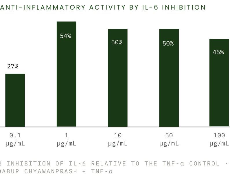
Mechanism
- 01TNF-α challenge to the endothelium
- 02IL-6 rises from ~20 to ~37 pg/mL
- 03DCP moderates IL-6 release
- 04Balanced endothelial inflammatory tone
Measured readouts
54%
Maximum inhibition, IL-6 ~17 pg/mL
50%
Inhibition vs TNF-α control
50%
Inhibition vs TNF-α control
45%
IL-6 close to untreated baseline
27%
IL-6 ~27 pg/mL
~20 → ~37
IL-6 pg/mL, untreated vs stimulated
Cellular benefits
- Supports heart and vascular health
- Supports a healthy blood vessel lining
- Helps maintain a balanced inflammatory response
- Helps protect vascular cells from inflammatory stress
Interpretation
The endothelium sits at the interface of inflammation and vascular health, so moderating its IL-6 response is the mechanistically relevant cardiovascular readout.
Supports cardiovascular health by maintaining a balanced inflammatory response.
DRF in-house endothelial cell assays.
Anti-inflammatory effect in human endothelial cells
To evaluate the cardiovascular health promoting potential of Dabur Chyawanprash (DCP) by anti-inflammatory effect in human endothelial cells (EA.hy926)
Human endothelial cells (EA.hy926)
Methodology
Dabur Chyawanprash (DCP) was dissolved in DMSO to make stock of 200 mg/ml, which was further diluted in serum free medium to achieve final concentrations for treatment.
Approximately 0.1 million (1 × 10) 5 endothelial cells were seeded in each well of a 24-well plate and incubated for 24 h to allow proper cell attachment and stabilization.
The cells were subsequently serum-starved for 24 h to minimize basal cellular activation and reduce the influence of serum-derived growth factors and cytokines, thereby providing a controlled background for assessment of the inflammatory response.
An inflammatory response was induced by exposing the cells to TNF-α (25 ng/mL), a well-established pro-inflammatory cytokine that activates endothelial inflammatory signaling and induces the secretion of downstream inflammatory mediators, including IL-6.
Cells were simultaneously treated with Dabur Chyawanprash (DCP) at concentrations ranging from 0.1–100 μg/mL in the presence of TNF-α and maintained for 48 h to evaluate the ability of DCP to modulate TNF-α-induced inflammatory activation.
Following treatment, the cell culture supernatants were collected and analyzed for secreted IL-6.
IL-6 levels were quantified using an ELISA, providing a quantitative measure of the inflammatory response induced by TNF-α.
The anti-inflammatory activity of DCP was determined by comparing IL-6 secretion in DCP-treated cells with the TNF-α-stimulated control cells. The reduction in IL-6 secretion was expressed as % inhibition relative to the TNF-α control.
A reduction in TNF-α-induced IL-6 secretion following DCP treatment indicates that DCP can attenuate inflammatory signaling in endothelial cells, supporting its potential to help maintain cellular homeostasis under inflammatory stress.
Thus, this model provides mechanistic in vitro evidence for the anti-inflammatory potential of DCP, particularly through modulation of IL-6, a key mediator associated with inflammatory responses.
% Anti-inflammatory activity was calculated as = {(Concentration of IL-6 in TNF-alpha stimulated cells alone – Concentration of IL-6 in DCP treated+TNF-alpha stimulated cells)/(Concentration of IL-6 in TNF-alpha stimulated cells alone }*100
Key biomarkers
This assay provides a biologically relevant in vitro model of inflammation by using TNF-α to induce inflammatory activation in endothelial cells. Measurement of IL-6 secretion by ELISA serves as a functional marker of this inflammatory response. Reduction of TNF-α-induced IL-6 levels following DCP treatment demonstrates its ability to attenuate inflammatory activation, providing mechanistic support for its potential to help maintain cellular and endothelial health under inflammatory stress.
Results
The results demonstrate that TNF-α markedly increased IL-6 secretion in endothelial cells, with IL-6 levels rising from approximately 20 pg/mL in untreated cells to ~37 pg/mL following TNF-α stimulation, confirming successful induction of an inflammatory response.
Treatment with Dabur Chyawanprash (DCP) significantly attenuated TNF-α-induced IL-6 secretion across the tested concentration range. At 0.1 μg/mL, DCP reduced IL-6 levels to approximately 27 pg/mL, corresponding to 27% inhibition compared with the TNF-α control. The strongest inhibition was observed at 1 μg/mL, where IL-6 levels decreased to approximately 17 pg/mL, representing 54% inhibition.
At 10 and 50 μg/mL, DCP maintained approximately 50% inhibition of IL-6, demonstrating a sustained anti-inflammatory effect. At 100 μg/mL, IL-6 levels remained close to the untreated baseline (~20 pg/mL), corresponding to approximately 45% inhibition relative to the TNF-α control.
Overall, DCP demonstrated a marked ability to suppress TNF-α-induced IL-6 secretion, with approximately 45–54% inhibition across 1–100 μg/mL. These findings provide in vitro evidence of the anti-inflammatory potential of DCP through attenuation of endothelial IL-6 release..
Scientific interpretation
The data indicate that DCP effectively modulates TNF-α-induced inflammatory activation in endothelial cells, as evidenced by a substantial reduction in IL-6 secretion. The maximum response was observed at 1 μg/mL, with 54% inhibition of IL-6, while approximately 45–50% inhibition was maintained at higher concentrations (10–100 μg/mL). This attenuation of a key pro-inflammatory cytokine suggests that DCP may interfere with inflammatory signaling triggered by TNF-α, thereby helping to limit endothelial inflammatory activation and support cellular homeostasis under inflammatory stress. Overall, the findings provide mechanistic in vitro evidence supporting the anti-inflammatory activity of DCP.
Anti-inflammatory activity by IL-6 inhibition
Scientific interpretation for consumer benefit claim
The inhibition of TNF-α-induced IL-6 secretion in endothelial cells demonstrates that DCP can attenuate an inflammatory response relevant to vascular endothelial health. Since excessive endothelial inflammation can contribute to vascular dysfunction and is associated with cardiovascular risk, the observed 45–54% reduction in IL-6 provides mechanistic in vitro evidence that DCP may help support healthy vascular function and maintain cardiovascular health by helping modulate inflammatory stress. This finding supports a potential consumer benefit of “supports cardiovascular health by helping maintain a balanced inflammatory response.”.
Potential consumer benefit claims:
- Supports cardiovascular health
- Supports healthy blood vessel function
- Helps maintain vascular health
- Helps support healthy endothelial function
- Helps maintain a balanced inflammatory response
- Helps protect vascular cells from inflammatory stress
Supports cellular health under inflammatory stress via cytokine inhibition in endothelial cells. IL-6 rose from approximately 20 pg/mL in untreated cells to ~37 pg/mL under TNF-α stimulation; bars give the percentage inhibition of IL-6 recorded at each DCP concentration.
Supporting neuronal health for cognitive function
Preclinical Evidence on Neuroprotective Activity and Neurite Outgrowth for Cognitive Wellness
Survival of human neuronal cells under MPP⁺ stress, neurite outgrowth in differentiated cells, and learning and memory in aged mice.
Model systemSH-SY5Y human neuronal cells; aged Swiss mice, CHY 1% and 2% w/w diet, 15 days
3 studies in this area
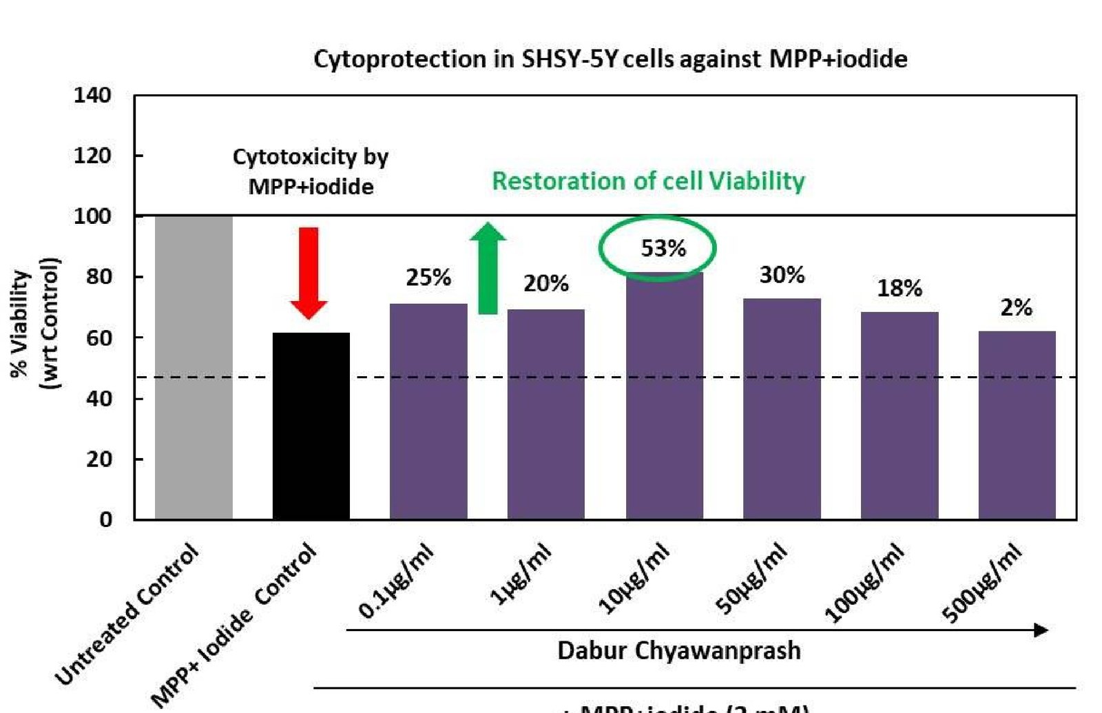 Study 1Cytoprotection in human neuronal cells (SH-SY5Y)To evaluate the neuronal health promoting potential of Dabur Chyawanprash (DCP) by cytoprotection in human neuronal cells (SH-SY5Y)
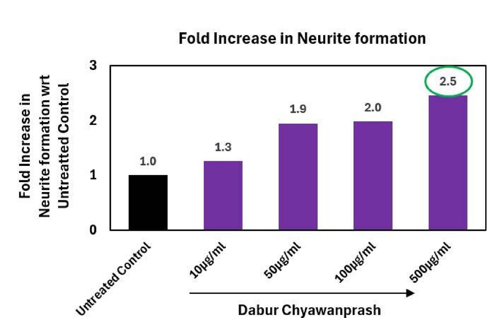 Study 2Increased Formation of Neurites in Human Neuronal Cells (SH-SY5Y)To evaluate the neuronal health promoting potential of Dabur
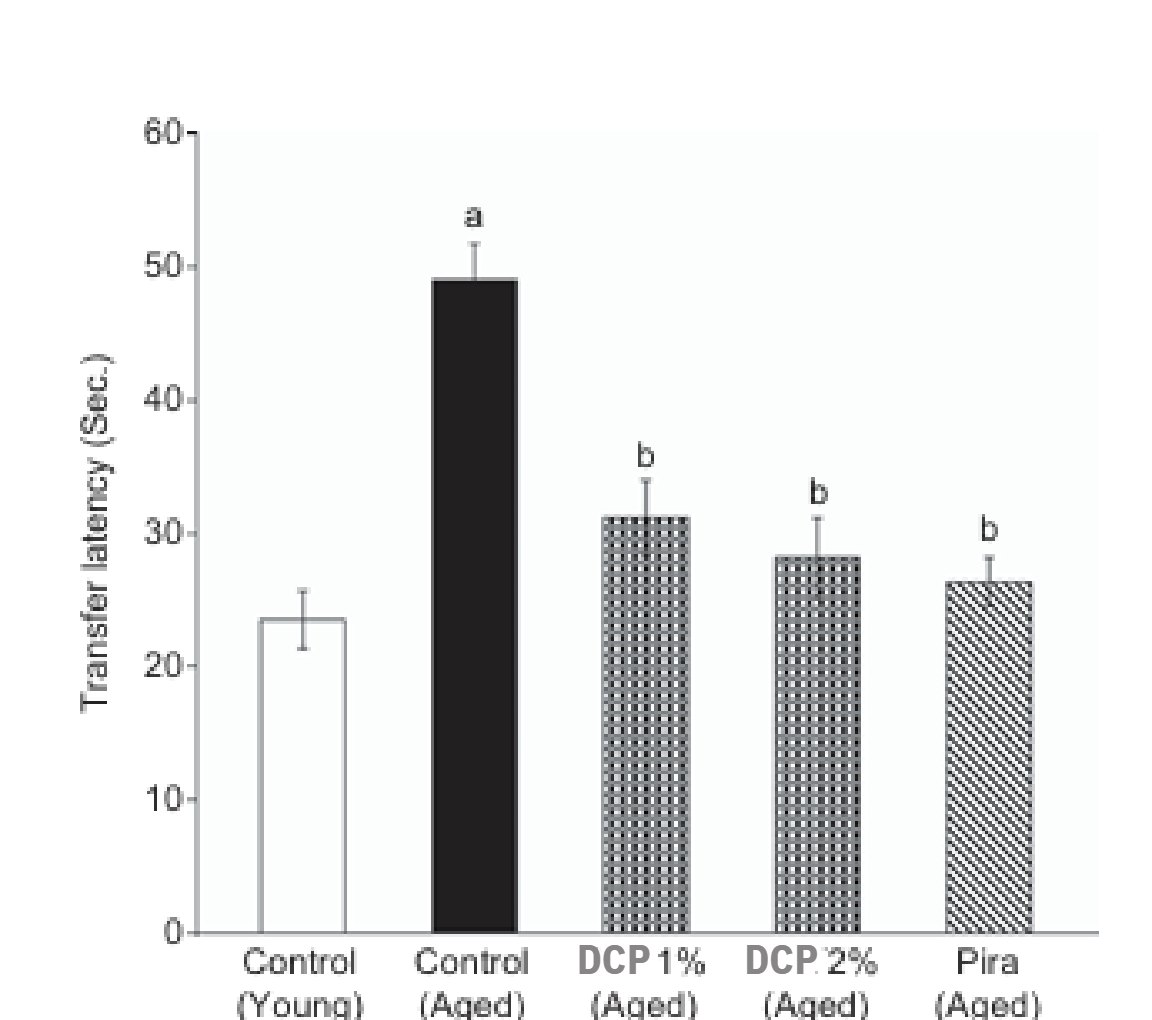 Study 3Beneficial Effect of Chyawanprash on Cognitive Function in Aged MiceTo investigate the beneficial effects of Dabur Chyawanprash (DCP) on cognitive function, learning and memory in aged mice, and to explore associated biochemical mechanisms.
Mechanism
- 01MPP⁺ and age-related oxidative stress
- 02Loss of viability and cholinergic decline
- 03DCP protects neurons, extends neurites, lowers AChE
- 04Preserved neuronal network and memory
Measured readouts
53%
At 10 µg/mL vs MPP⁺ (viability ~62%)
2.5×
At 500 µg/mL (1.3×, 1.9×, 2.0× at 10–100)
54.2 → 35.4 s
Morris water maze Day 4, CHY 2%
28.2 → 51.2 s
Memory retrieval, CHY 2%
↓ · ↑ GSH
Brain biomarkers, CHY 1% and 2%
No change
Effects not due to general activity
Cellular benefits
- Supports brain health
- Supports learning and memory (aged-animal model)
- Protects neurons from oxidative stress
- Supports healthy neuronal development
Interpretation
Survival alone is not sufficient for cognition. Neurite outgrowth and the aged-mouse memory data show that connectivity and function are also supported.
Supports neuronal health and healthy cognitive function.
DRF in-house neuronal cell assays and published aged-mouse study.
Cytoprotection in human neuronal cells (SH-SY5Y)
To evaluate the neuronal health promoting potential of Dabur Chyawanprash (DCP) by cytoprotection in human neuronal cells (SH-SY5Y)
Human neuronal cells (SH-SY5Y)
Methodology
Dabur Chyawanprash (DCP) was dissolved in DMSO to make stock of 200 mg/ml, which was further diluted in serum free medium to achieve final concentrations for treatment.
10,000 neuronal cells were seeded in each well of a 96-well plate and incubated for 24 hours to allow proper cell attachment and stabilization.
Cells were subsequently treated with Dabur Chyawanprash (DCP) at concentrations ranging from 0.1–500 μg/mL for 24 hours to evaluate its potential protective effect on neuronal cells.
Following DCP treatment, neuronal stress was induced by exposing the cells to 2 mM MPP⁺ iodide overnight. MPP⁺ is a wellestablished neurotoxic agent that induces mitochondrial dysfunction, oxidative stress and loss of neuronal cell viability, thereby serving as an in vitro model of chemically induced neuronal injury.
After MPP⁺ exposure, cellular viability was assessed using the MTT assay, which measures cellular metabolic activity as an indicator of viable cells. MPP⁺-induced cellular damage was reflected by a reduction in MTT-derived viability compared with untreated cells.
The cytoprotective potential of DCP was determined by comparing the viability of DCP + MPP⁺-treated cells with that of the MPP⁺- exposed control cells. An increase in viability following DCP treatment indicated attenuation of MPP⁺-induced cellular damage.
The percentage cytoprotection was calculated relative to the MPP⁺-induced injury control, thereby determining the extent to which DCP restored neuronal cell viability following neurotoxic stress.
Overall, the model was designed to determine whether DCP can preserve neuronal cell viability and provide cellular protection against MPP⁺-induced neurotoxic/oxidative stress, supporting its potential role in maintaining neuronal cellular health.
The percentage cell viability was calculated as = 100-%cytotoxicity. % Cytotoxicity was calculated as – ((Absorbance of untreated – Absorbance of treated)/(Absorbance of Untreated))*100. % Cytoprotection was calculated as = {(Absorbance of DCP-treated – Absorbance of MPP+ damaged cells)/(Absorbance of untreated – Absorbance of MPP+ damaged cells}*100
Restoration of cell viability as compared to MPP+ induced control, was considered indicative of cytoprotective response.
Key biomarkers
The MPP⁺-induced neuronal cytotoxicity model is relevant to neuronal health as it mimics oxidative and mitochondrial stress associated with neuronal injury. MPP⁺ reduces cellular viability through mitochondrial dysfunction and oxidative damage. Therefore, restoration of neuronal cell viability by DCP indicates its potential to enhance neuronal cellular resilience and protect neuronal cells against stress-induced damage, supporting a potential role in maintaining neuronal health.
Results
MPP⁺ iodide exposure reduced SH-SY5Y neuronal cell viability to approximately 62% of the untreated control, confirming induction of significant cellular stress.
Treatment with DCP showed restoration of neuronal cell viability at most concentrations tested. The highest cytoprotective response was observed at 10 μg/mL, with 53% restoration, followed by 30% at 50 μg/mL and 25% at 0.1 μg/mL. Lower protection was observed at 1 and 100 μg/mL, while 500 μg/mL showed minimal protection (2%).
Overall, the results indicate that DCP can attenuate MPP⁺-induced neuronal cytotoxicity and support neuronal cell survival under stress conditions, with 10 μg/mL showing the most pronounced protective effect.
Scientific interpretation
The results demonstrate that MPP⁺ iodide induced substantial cytotoxic stress in SH-SY5Y neuronal cells, as evidenced by the reduction in cell viability to approximately 62% of the untreated control. DCP treatment attenuated this MPP⁺- induced loss of viability, with the maximum cytoprotective effect observed at 10 μg/mL (53% cytoprotection). The protective response at several concentrations indicates that DCP may enhance neuronal cellular resilience against MPP⁺- induced oxidative/mitochondrial stress. The reduced effect at higher concentrations suggests that the response is not strictly concentration-dependent, highlighting 10 μg/mL as the most effective concentration under the conditions tested.
Scientific interpretation for consumer benefit claim
The findings indicate that DCP can help protect neuronal cells from stress-induced damage and support maintenance of neuronal cell viability. The observed restoration of viability following MPP⁺-induced injury, particularly the 53% cytoprotection at 10 μg/mL, supports a potential consumer benefit related to maintaining neuronal cellular health and resilience against oxidative/ cellular stress. This provides mechanistic support for positioning DCP as contributing to neuronal health and protection against cellular stress, rather than making disease-prevention or treatment claims.
Potential consumer benefit claims:
- Supports neuronal health and cellular resilience
- Helps protect neuronal cells against cellular stress
- Supports the natural protection of neuronal cells from oxidative stress
- Helps maintain healthy neuronal cell viability
- Supports neuronal cells against stress-induced damage
- Helps maintain neuronal cellular health and function
- Provides cellular protection against stress-related neuronal damage
- Supports the resilience of neuronal cells under oxidative stress conditions
Increased Formation of Neurites in Human Neuronal Cells (SH-SY5Y)
To evaluate the neuronal health promoting potential of Dabur
Chyawanprash (DCP) by increased formation of neurites in human neuronal cells (SH-SY5Y)
Human neuronal cells (SH-SY5Y)
Methodology
Dabur Chyawanprash (DCP) was dissolved in DMSO to make stock of 200 mg/ml, which was further diluted in serum free medium to achieve final concentrations for treatment.
Day 0: Approximately 10,000 SH-SY5Y neuronal cells were seeded in each well of a 12-well plate in DMEM supplemented with 10% FBS and incubated for 24 hours to allow cell attachment and recovery.
Day 1: Following overnight incubation, neuronal differentiation was initiated by replacing the growth medium with differentiation medium containing 1 μM retinoic acid (RA). Cells were maintained under these differentiation conditions for 6 days, with the medium replaced every 2 days to maintain appropriate nutrient and differentiation conditions.
Day 6: After establishment of the differentiated neuronal phenotype, cells were treated with Dabur Chyawanprash (DCP) at concentrations ranging from 0.1–500 μg/mL for 48 hours in the same differentiation medium. Cells maintained in differentiation medium without DCP served as the untreated control.
Following treatment, cellular morphology and neurite development were assessed using phase-contrast microscopy. Representative images were captured and the number of neurites extending from differentiated neuronal cells was quantified.
The effect of DCP on neurite development was expressed as the percentage increase in neurite formation relative to the untreated differentiated control.
This experimental design specifically evaluates whether DCP can further promote neurite formation in an already differentiated neuronal model, providing evidence for its potential effects on neuronal maturation and structural development.
Fold increase in neurite formation = {(No of neurites in DCP-treated cells – No of neurites in Untreated cells)/No of neurites in untreated cells)}*100
Key biomarkers
This assay is relevant to neuronal health because neurite outgrowth is a key feature of neuronal differentiation, maturation and development of functional neuronal networks. Increased neurite formation indicates enhanced neuronal growth and structural development. Therefore, the assay helps determine whether DCP can support neuronal differentiation and neurite development, providing mechanistic evidence for its potential role in maintaining neuronal health, connectivity and cellular function.
Results
DCP treatment produced a progressive increase in neurite formation in retinoic acid-differentiated SH-SY5Y neuronal cells compared with the untreated differentiated control.
Neurite formation increased from 1.0-fold in the control to 1.3-fold at 10 μg/mL, 1.9-fold at 50 μg/mL, and 2.0-fold at 100 μg/mL. The strongest response was observed at 500 μg/mL, with a 2.5-fold increase in neurite formation compared with the untreated control.
Overall, the results demonstrate a concentration-responsive enhancement of neurite development by DCP, suggesting that DCP may support neuronal maturation and structural development, with the most pronounced effect observed at 500 μg/mL
Scientific interpretation
The results indicate that DCP enhanced neurite formation in retinoic acid-differentiated SH-SY5Y neuronal cells, with the response increasing progressively from 1.3-fold at 10 μg/mL to 2.5-fold at 500 μg/mL. This suggests that DCP may promote neuronal maturation and structural development, as neurite extension is an important feature of neuronal differentiation and establishment of neuronal connections.
The concentration-responsive increase provides mechanistic evidence that DCP may support neuronal growth, connectivity and cellular health.
Scientific interpretation for consumer benefit claim
The findings provide mechanistic evidence that DCP may support neuronal structural health and development. In retinoic acid-differentiated SH-SY5Y cells, DCP increased neurite formation by up to 2.5-fold at 500 μg/mL compared with the untreated differentiated control, with progressive enhancement across the tested concentrations. Neurite formation is an important feature of neuronal maturation and is associated with the development of cellular extensions required for neuronal communication and network formation. Therefore, the observed increase suggests that DCP may support neuronal growth, maturation and structural connectivity, contributing to maintenance of healthy neuronal function. For consumer communication, the data can support broader claims such as “supports neuronal health,” “supports healthy neuronal development,” and “helps maintain neuronal connectivity,”.
Potential consumer benefit claims:
- Supports neuronal health and function
- Supports healthy neuronal development and maturation
- Helps support neurite growth and development
- Supports healthy neuronal connectivity
- Helps maintain the structural health of neurons
- Supports neuronal growth and cellular resilience
- Helps maintain healthy neuronal communication
- Supports brain cell health and development
- Helps support the natural growth and connectivity of neuronal cells
Beneficial Effect of Chyawanprash on Cognitive Function in Aged Mice
To investigate the beneficial effects of Dabur Chyawanprash (DCP) on cognitive function, learning and memory in aged mice, and to explore associated biochemical mechanisms.
Male Swiss mice. Young mice were 3–4 months old (approximately 20 g) and aged mice were 12–15 months old (approximately 35 g). Each treatment group comprised a minimum of six animals.
Method
Animals were acclimatized for at least 5 days before behavioral experiments.
Dabur Chyawanprash was administered orally by mixing with the standard diet at two concentrations: DCP 1% w/w and CHY 2% w/w.
Treatment continued for 15 successive days. Young and aged control groups received standard diet without DCP.
Piracetam 400 mg/kg i.p. was the positive control for behavioral tests; donepezil 0.1 mg/kg i.p. was the standard drug for biochemical estimations.
After treatment, animals were evaluated using the Morris water maze and elevated plus maze. Brain tissue was collected for biochemical estimation of AChE, TBARS and GSH. Locomotor activity was also measured.
Study at a glance
SPECIES Male Swiss mice
DOSE DCP 1% w/w and DCP 2% w/w, in standard diet
GROUP SIZE Minimum six animals per group
STANDARD DRUG Donepezil 0.1 mg/kg i.p. (biochemical)
Key biomarkers
Behavioral: Morris water maze escape latency time (ELT) and time spent in target quadrant (TSTQ); elevated plus maze transfer latency (TL).
Biochemical: brain acetylcholinesterase (AChE), thiobarbituric acid reactive substances (TBARS) and reduced glutathione (GSH).
Control for confounding: spontaneous locomotor activity.
Design note
A 15-day dietary administration model in naturally aged animals, with both a nootropic positive control and a cholinesteraseinhibitor standard, allowing behavioral and biochemical outcomes to be read against age-matched controls.
AGE GROUPS Young 3–4 months (~20 g) · Aged 12–15 months (~35 g)
DURATION 15 successive days, oral
POSITIVE CONTROL Piracetam 400 mg/kg i.p. (behavioral)
BEHAVIORAL TESTS Morris water maze · elevated plus maze
Results
Morris water maze: learning and memory retrieval a · escape latency, day 4
Aged control mice showed a markedly higher Day 4 escape latency than young control mice, indicating age-associated learning impairment. DCP treatment reduced Day 4 escape latency in aged mice, and the reductions produced by DCP 1% and DCP 2% indicated improved learning, with the 2% treatment showing a greater reduction than the 1% treatment.
B · time spent in target quadrant
Aged control mice also spent significantly less time in the target quadrant than young controls. DCP significantly increased the time spent in the target quadrant in aged mice, indicating improved memory retrieval; the 2% group showed a stronger effect than the 1% group.
C · elevated plus maze, transfer latency
Aged mice showed higher transfer latency than young mice, demonstrating impaired memory. Treatment with both DCP 1% and DCP 2% significantly decreased Day 16 transfer latency in aged mice. Piracetam produced a similar positive effect.DCP 2% produced a greater magnitude of improvement tha DCP 1% across the water-maze outcomes.
Aged control 54.2 ± 2.6
DCP 1% 43.8 ± 1.1 35.4 ± 2.3 DCP 2%
Piracetam 21.4 ± 1.7
Mean ± SEM, seconds.
Young control 73.4 ± 3.6
Aged control 28.2 ± 1.9
DCP 1% 39.1 ± 1.8
DCP 2% 51.2 ± 4.6
Piracetam 54.3 ± 2.8
Mean ± SEM, seconds.
Cholinergic and oxidative-stress biomarkers d · brain acetylcholinesterase
Aged mice exhibited significantly increased brain AChE activity compared with young mice. Treatment with both DCP 1% and DCP 2% significantly reduced AChE activity. Donepezil produced a similar reduction and served as the standard biochemical control.
E · brain TBARS and GSH
Aged mice had higher TBARS levels, indicating increased oxidative stress, and lower GSH levels, indicating reduced antioxidant status. DCP 1% and DCP 2% treatment reduced brain TBARS and increased GSH levels in aged mice. These effects were also observed with donepezil.
F · locomotor activity
DCP at both 1% and 2% w/w did not produce a significant change in locomotor activity compared with control animals, suggesting that the observed cognitive effects were not simply due to changes in general locomotor activity.
G · safety and general parameters
Administration of DCP for 15 days did not significantly alter feed consumption or body weight. All animals survived in each treatment group, supporting the safety of the treatment under the conditions of the study.
The study demonstrated that ageing was associated with impaired learning and memory, increased brain AChE activity, increased oxidative stress, and decreased GSH levels. Fifteen days of Dabur Chyawanprash treatment improved behavioral measures of learning and memory in aged mice while reducing AChE activity and oxidative stress and increasing GSH levels.
Dabur Chyawanprash may act as a memory enhancer in aged animals through multiple beneficial effects:
Improvement of learning ability
Improvement of memory retrieval and retention
Reduction of brain acetylcholinesterase activity
Enhancement of cholinergic transmission
Reduction of oxidative stress, reflected by lower TBARS
Enhancement of antioxidant defence, reflected by increased GSH
It was concluded that Dabur Chyawanprash may be useful for improving memory in aged animals because of its multifactorial, antioxidant and pro-cholinergic effects. Further investigation is required to determine the involvement of other neurotransmitter systems.
Scientific interpretation
The study provides convergent behavioral and biochemical evidence for a beneficial effect of Dabur Chyawanprash on age-associated cognitive impairment in mice.
Improvement in learning and memory was demonstrated across two behavioral paradigms. Reduced escape latency in the Morris water maze indicates improved learning, while increased time in the target quadrant indicates improved spatial memory retrieval. Reduced transfer latency in the elevated plus maze supports improved memory retention.
The reduction in brain AChE activity provides a potential cholinergic mechanism, which may facilitate cholinergic neurotransmission and contribute to the improved memory.
The reduction in TBARS together with an increase in GSH suggests an improvement in brain antioxidant status and reduced oxidative stress.
The effect is characterized as multifactorial, potentially involving both pro-cholinergic and antioxidant mechanisms. The 2% treatment generally produced a greater magnitude of improvement than 1% for the reported Morris water maze outcomes.
Scientific interpretation for consumer benefit claim
Supports cognitive performance, learning, memory retrieval and memory retention in an aged-animal model, with associated changes in cholinergic and antioxidant biomarkers.
Bansal N, Parle M. Beneficial effect of Chyawanprash on cognitive function in aged mice. Pharmaceutical Biology. 2011;49(1):2–8.
DOI: 10.3109/13880209.2010.489904.
Driving bone-forming cell activity
Substantiated by Osteoblast Proliferation Evidence for Bone Formation & Strength
Osteoblasts are the cells that lay down bone matrix. DCP increased their proliferation in a concentration-dependent manner over 72 hours.
Model systemMG63 human osteoblasts, 72 h, versus untreated control
1 study in this area
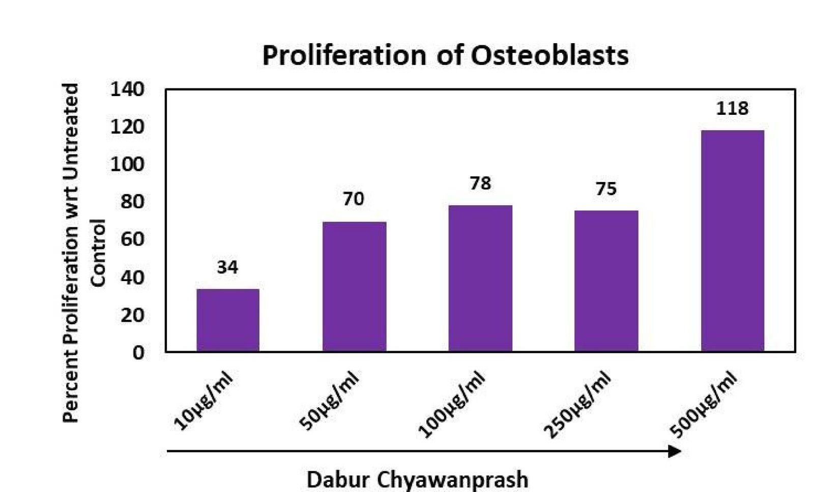
Mechanism
- 01DCP bioactive phytoconstituents
- 02Uptake by osteoblasts and metabolic activity
- 03Increased proliferation
- 04Support for bone-forming activity
Measured readouts
118%
Of untreated control at 500 µg/mL
+18%
Highest concentration exceeds control
78%
Proliferation vs untreated control
75%
Sustained response
70%
Proliferation vs untreated control
34%
Proliferation vs untreated control
Cellular benefits
- Supports bone health
- Supports healthy osteoblast activity
- Helps maintain strong bones
- Supports natural bone renewal
Interpretation
Proliferation rose with concentration and exceeded baseline at 500 µg/mL. Because osteoblasts build bone matrix, this is an early functional signal for bone formation.
Supports bone health and healthy bone-forming cell activity.
DRF in-house MG63 osteoblast cells assay.
Proliferation of Human Osteoblasts (MG63)
To evaluate the bone health promoting potential of Dabur Chyawanprash (DCP) by increase in proliferation of human osteoblasts (MG63)
Human osteoblasts (MG63)
Methodology
Dabur Chyawanprash (DCP) was dissolved in DMSO to make stock of 200 mg/ml, which was further diluted in serum free medium to achieve final concentrations for treatment.
1,000 osteoblast cells were seeded per well in a 96-well plate and incubated for 24 h to allow cell attachment and stabilization.
Cells were subsequently serum-starved for 24 h in DMEM containing 0.1% FBS to minimize serum-driven proliferation and establish a controlled baseline for evaluating the direct effect of DCP on osteoblast proliferation.
The cells were then treated with Dabur Chyawanprash (DCP) at concentrations ranging from 10–500 μg/mL and maintained for 72 h to assess its ability to stimulate osteoblast growth and proliferation.
Following treatment, cellular proliferation/metabolic activity was evaluated using the MTT assay. The assay measures the ability of viable metabolically active cells to reduce MTT to formazan, with the resulting absorbance serving as an indirect measure of viable cell number.
The proliferative response of DCP-treated osteoblasts was expressed as % proliferation relative to the untreated control, which was considered the baseline (100%).
An increase in MTT-derived viability/metabolic activity compared with the untreated control indicates an increase in osteoblast proliferation, suggesting that DCP may support osteoblast cellular growth and activity.
Thus, this assay provides in vitro biological evidence for the potential osteoblast-supportive activity of DCP, which may be relevant to maintaining bone-forming cellular function and bone health.
% increase in osteoblasts proliferation was calculated as = {(Absorbance of DCP treated cells – Absorbance of Untreated cells)/(Absorbance of Untreated cells }*100
Key biomarkers
This osteoblast proliferation model is biologically relevant because osteoblasts are the primary bone-forming cells responsible for bone matrix production and mineralization. Assessing the ability of DCP to enhance osteoblast proliferation provides an early functional indication of its potential to support bone-forming cellular activity and bone tissue maintenance. The 72-hour treatment and MTT-based assessment provide a quantitative measure of changes in cellular metabolic activity and viable cell number, thereby offering in vitro mechanistic support for a potential bone-health benefit.
Results
The results demonstrate that Dabur Chyawanprash (DCP) enhanced osteoblast proliferation in a concentration-dependent manner across the tested concentration range. At 10 μg/mL, osteoblast proliferation was approximately 34% of the untreated control, increasing substantially to 70% at 50 μg/mL and 78% at 100 μg/mL. At 250 μg/mL, proliferation remained high at 75%, indicating a sustained cellular response.
The maximum proliferative response was observed at 500 μg/mL, reaching 118% relative to the untreated control, representing an approximately 18% increase above the untreated baseline.
Overall, DCP demonstrated a progressive enhancement of osteoblast cellular proliferation with increasing concentration, with the highest concentration restoring/enhancing proliferation beyond the untreated control level. These findings suggest that DCP may support osteoblast growth and bone-forming cellular activity, providing preliminary in vitro mechanistic evidence for its potential role in supporting bone health.
Scientific interpretation
The results indicate that DCP positively modulates osteoblast proliferation, with the response increasing from 34% at 10 μg/mL to 118% at 500 μg/mL relative to the untreated control. The highest concentration increased proliferation above the untreated baseline, suggesting enhanced osteoblast cellular activity. As osteoblasts are responsible for bone formation and matrix deposition, these findings provide mechanistic in vitro evidence that DCP may support osteoblast function and contribute to the maintenance of bone health.
Scientific interpretation for consumer benefit claim
The enhanced proliferation of osteoblasts following DCP treatment, reaching 118% of the untreated control at 500 μg/mL, indicates a potential stimulatory effect on boneforming cellular activity. Since osteoblasts play a central role in bone formation and maintenance, these findings provide mechanistic in vitro support for DCP’s potential to support bone health by promoting healthy osteoblast activity and cellular renewal. This may translate into a consumer benefit of “supports healthy bones and bone-forming activity.”
Potential consumer benefit claims:
- Supports healthy bone formation
- Helps maintain strong and healthy bones
- Supports healthy osteoblast activity
- Helps support bone-forming cellular activity
- Supports maintenance of bone tissue
- Helps promote healthy bone-cell function
- Supports bone strength and structural health
Three mechanisms against inflammaging
Preclinical Evidence on Beta-galactosidase Inhibition, GPCR-Mediated Pathways & Telomerase Activation for Cellular Wellness & Longevity
Inflammaging, the chronic low-grade inflammation of ageing tissue, is addressed on three fronts: telomerase activity, senescence-associated β-galactosidase and CXCR3 chemokine signalling.
Model systemHuman PBMCs; CXCR3-overexpressing HeLa reporter cells
3 studies in this area
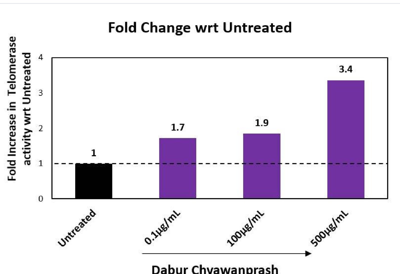 Study 1Telomerase Activation in Human PBMCsTo evaluate the effect of Dabur Chyawanprash (DCP) on Telomerase activation in human Peripheral Blood Mononuclear Cell (PBMCs)
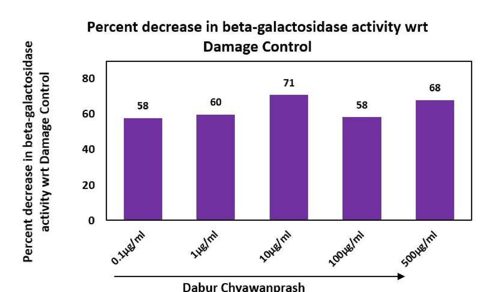 Study 2Inhibition of Beta-Galactosidase in Human PBMCsTo evaluate the effect of Dabur Chyawanprash (DCP) on inhibition of Beta-Galactosidase in human Peripheral Blood Mononuclear Cell (PBMCs)
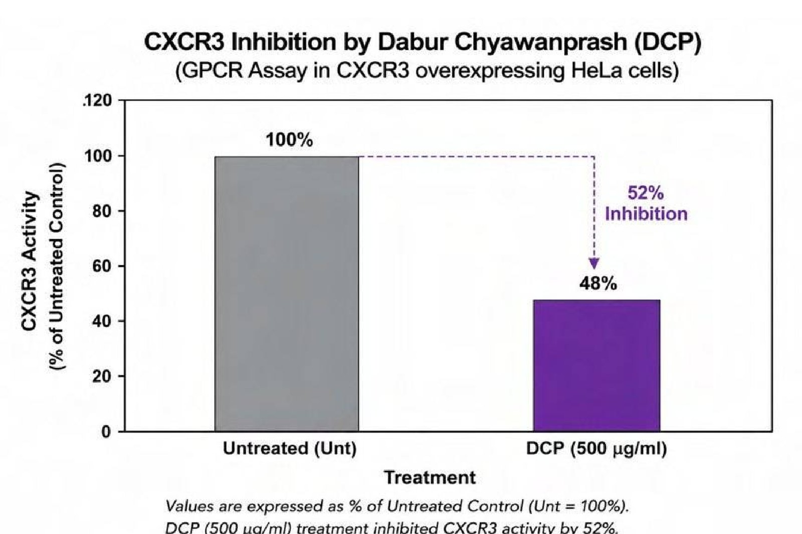 Study 3Inhibition of CXCR3 (GPCR) in Receptor Overexpressing HeLa Cell LineTo evaluate the anti-inflammatory effect of Dabur Chyawanprash (DCP) via inhibition of C-X-C chemokine receptor 3; CXCR3 (GPCR) in receptor overexpressing Hela cell line
Mechanism
- 01Chronic low-grade inflammatory signalling
- 02Cellular senescence and rising oxidative load
- 03DCP raises telomerase, lowers β-gal, blocks CXCR3
- 04Restored cellular vitality
Measured readouts
3.4×
At 500 µg/mL vs untreated PBMCs
1.7× · 1.9×
At 0.1 and 100 µg/mL
71%
Maximum at 10 µg/mL, t-BHP model
58–71%
Across 0.1–500 µg/mL
52%
Receptor inhibition, GPCR reporter assay
t-BHP 5 mM
3 h oxidative challenge
Cellular benefits
- Supports healthy ageing
- Supports cellular vitality
- Helps maintain a balanced inflammatory response
- Supports natural cellular renewal and maintenance
Interpretation
Inflammation, oxidation and senescence reinforce one another, so acting on all three simultaneously is more consequential than any single readout suggests.
Supports cellular vitality and healthy ageing.
DRF in-house PBMC assays and external GPCR profiling.
Telomerase Activation in Human PBMCs
To evaluate the effect of Dabur Chyawanprash (DCP) on Telomerase activation in human Peripheral Blood Mononuclear Cell (PBMCs)
Human Peripheral Blood Mononuclear Cell (PBMCs)
Methodology
Dabur Chyawanprash (DCP) was dissolved in DMSO to make stock of 200 mg/ml, which was further diluted in serum free medium to achieve final concentrations for treatment.
The assay was designed to evaluate the effect of Dabur Chyawanprash (DCP) on telomerase activity in human peripheral blood mononuclear cells (PBMCs). Telomerase is an enzyme complex that maintains telomere length and is associated with cellular replicative capacity and longevity.
Day 1 – Cell seeding: Human PBMCs were seeded at a density of 1.5 × 10⁶ cells per well in 6-well plates and maintained for 24 h in RPMI-1640 medium supplemented with 0.5% FBS. The reducedserum condition was used to provide a controlled cellular environment prior to treatment.
Following the initial incubation, PBMCs were treated with DCP at concentrations ranging from 0.1–500 μg/mL. Cells were maintained in RPMI-1640 containing 0.5% FBS for 48 h. An untreated control group was maintained under identical conditions without DCP treatment.
Day 3 – Cell lysate preparation: After 48 h of treatment, cells were harvested and lysates were prepared under appropriate conditions for assessment of telomerase activity.
Quantification of telomerase activity: Relative telomerase activity was determined using a qPCR-based assay, in which the telomerase-dependent amplification signal was quantified and compared with the untreated control.
Data analysis: Telomerase activity was expressed as fold change relative to the untreated control, which was assigned a value of 1.0. The fold increase was calculated to determine the extent to which DCP treatment enhanced telomerase activity compared with baseline cellular levels.
Fold increase in Telomerase activity = {(Activity in DCP-treated cells-Activity in Untreated cells)/ Activity in Untreated cells}*100
Key biomarkers
The PBMC model provides a physiologically relevant human-cell system for evaluating modulation of telomerase activity. An increase in relative telomerase activity following DCP treatment indicates that the formulation can stimulate telomerase-associated cellular maintenance mechanisms in human immune cells. Such activity may be relevant to cellular longevity, maintenance of replicative potential and protection against telomere-associated cellular senescence.
Results
The graph demonstrates that Dabur Chyawanprash (DCP) enhanced relative telomerase activity in human PBMCs in a concentration-dependent manner compared with the untreated control.
Untreated control: Telomerase activity was normalized to 1.0-fold, representing the baseline level.
DCP 0.1 μg/mL: Telomerase activity increased to 1.7-fold, corresponding to approximately a 70% increase over untreated cells.
DCP 100 μg/mL: Telomerase activity increased further to 1.9-fold, representing approximately a 90% increase over baseline.
DCP 500 μg/mL: The highest response was observed, with telomerase activity reaching 3.4-fold, equivalent to approximately a 240% increase over untreated control.
Scientific interpretation
The progressive increase from 1.7 1.9 3.4-fold with increasing DCP concentrations indicates a dose-responsive stimulation of telomerase activity in PBMCs. The maximum response at 500 μg/mL suggests that DCP can substantially enhance telomerase-associated cellular maintenance mechanisms in human immune cells.
Scientific interpretation for consumer benefit claim
The results demonstrate that Dabur Chyawanprash (DCP) enhanced telomerase activity in human PBMCs, with the response increasing from 1.7-fold at 0.1 μg/mL to 3.4-fold at 500 μg/mL compared with untreated cells. The progressive increase with increasing DCP concentration indicates a doseresponsive stimulation of telomerase activity. Telomerase is a key cellular enzyme involved in telomere maintenance and preservation of cellular replicative potential. Therefore, the observed enhancement of telomerase activity provides mechanistic evidence that DCP may support cellular resilience, healthy cellular aging and maintenance of cellular longevity-related processes. Telomere maintenance Cellular vitality Healthy cellular aging Inflammatory balance Healthy aging
Potential consumer benefit claims:
- Supports cellular vitality
- Supports healthy cellular aging
- Helps maintain cellular health and resilience
- Supports natural cellular renewal and maintenance
Inhibition of Beta-Galactosidase in Human PBMCs
To evaluate the effect of Dabur Chyawanprash (DCP) on inhibition of Beta-Galactosidase in human Peripheral Blood Mononuclear Cell (PBMCs)
Human Peripheral Blood Mononuclear Cell (PBMCs)
Methodology
Dabur Chyawanprash (DCP) was dissolved in DMSO to make stock of 200 mg/ml, which was further diluted in serum free medium to achieve final concentrations for treatment.
This assay was designed to evaluate the protective effect of Dabur Chyawanprash (DCP) against oxidative stress–induced cellular senescence. β-Galactosidase activity is widely used as a marker associated with the senescence-associated phenotype, particularly when measured under conditions optimized for senescence-associated β-galactosidase detection.
Day 0 – Cell seeding: Approximately 1 × 10⁶ cells were seeded in each well of a 6-well plate and cultured for 24 h in DMEM supplemented with 10% FBS to allow adequate cell attachment and recovery.
Day 1 – Serum starvation: Cells were subjected to serum starvation for 24 h using DMEM containing 0% FBS. This step was performed to establish a controlled cellular environment and minimize the influence of serum-derived growth factors before treatment.
Day 2 – DCP treatment: Cells were treated with DCP at concentrations ranging from 0.1–500 μg/mL for 24 h in serum-free DMEM. An appropriate untreated/control group was maintained in parallel.
Day 3 – Oxidative stress induction: Following DCP trea tment, cells were exposed to tert-butyl hydroperoxide (t-BHP; 5 mM) for 3 h to induce oxidative cellular damage and a senescence-associated response. t-BHP acts as a potent oxidative stressor and can generate intracellular oxidative stress, leading to cellular damage and activation of pathways associated with senescence.
β-Galactosidase measurement: Following oxidative challenge, cell lysates were prepared and β-galactosidase activity was quantified using the fluorescence mode of a spectrophotometer. The fluorescence signal served as a quantitative measure of β-galactosidase activity.
Data analysis: The response obtained in the damage control (t-BHP-treated cells without DCP) was used as the reference for calculating the percentage decrease in β-galactosidase activity following DCP treatment.
% Inhibition in Beta-Gal activity = {(Activity in damaged cells-Activity in DCP-treated cells)/Activity in damaged cells}*100
Key biomarkers
The t-BHP–induced senescence model is relevant to cellular vitality because it evaluates the ability of DCP to help cells withstand oxidative stress–associated loss of cellular function. Oxidative stress can accelerate cellular damage and promote a senescence-associated phenotype, reflected in increased β-galactosidase activity. Therefore, the observed reduction in β-galactosidase activity following DCP treatment suggests that DCP may help attenuate oxidative stress–associated cellular senescence and preserve cellular fitness.
Mechanistic relevance
Oxidative stress cellular damage senescence-associated phenotype reduced cellular vitality. DCP attenuation of senescence-associated response preservation of cellular fitness/vitality.
Results
The results demonstrate that Dabur Chyawanprash (DCP substantially reduced β-galactosidase activity in the oxidative damage model compared with the damage control. Since β-galactosidase is used as a senescence-associated marker, its reduction suggests attenuation of the oxidative stress–induced senescence response. 0.1 μg/mL: DCP produced a 58% decrease in β-galactosidase activity.
1 μg/mL: A 60% decrease was observed.
10 μg/mL: The maximum response was observed, with a 71% decrease in β-galactosidase activity.
100 μg/mL: β-galactosidase activity was reduced by 58%.
500 μg/mL: A 68% decrease was observed.
Thus, DCP demonstrated a consistently strong reduction of approximately 58–71% across the entire tested concentration range, with the highest effect at 10 μg/mL
Scientific interpretation
The substantial reduction in β-galactosidase activity indicates that DCP attenuated the cellular senescence-associated response induced by oxidative stress. The effect was evident even at the lowest tested concentration, suggesting that DCP may enhance the ability of cells to maintain cellular fitness under oxidative challenge. These findings support the concept that DCP can help preserve cellular vitality and resilience under oxidative stress by reducing a key marker associated with cellular senescence.
Scientific interpretation for consumer benefit claim
Oxidative stress is an important contributor to cellular dysfunction and loss of cellular vitality. By reducing the oxidative stress–associated increase in β-galactosidase activity, DCP demonstrates potential to support cellular resilience and preserve cellular vitality under stress conditions. The maximum 71% reduction at 10 μg/mL provides strong mechanistic evidence for an effect on cellular senescence-associated pathways. Although the response was not strictly dose-dependent, the consistently high reduction across concentrations suggests a robust biological effect within the tested range.
Potential consumer benefit claims:
- Supports cellular vitality
- Helps maintain cellular resilience against oxidative stress
- Supports cellular health and function
- Helps protect cells from oxidative stress–associated damage
- Supports natural cellular maintenance
Inhibition of CXCR3 (GPCR) in Receptor Overexpressing HeLa Cell Line
To evaluate the anti-inflammatory effect of Dabur Chyawanprash (DCP) via inhibition of C-X-C chemokine receptor 3; CXCR3 (GPCR) in receptor overexpressing Hela cell line
Human receptor overexpressing Hela cell line
Methodology
The comprehensive GPCR profiling was performed in Tanso Biosciences, Inc., Japan, using Tanso™ Biosciences’ proprietary high-throughput GPCR reporter assay platform.
Cell handling
Proprietary reporter cells (3) are maintained in DMEM supplemented with 10% FCS (pH 7.4) and penicillin-streptomycin according to standard procedures.
Cells detached by trypsin-EDTA were suspended in DMEM containing 10% dialyzed FCS (pH 7.4).
Suspended cells were seeded into white-walled 96-well microplates at a density of 24000 cells per well.
Cells were allowed to adhere to microplates in a 5% CO₂ incubator at 37°C and cultured overnight.
Transient transfection
Transfection was performed using FuGENE6 transfection reagent.
Cells were transiently transfected in a 96-well microplate with a mixture of plasmids encoding GPCR, Gα protein, and luciferasereporter (total 100 ng per well).
Transfected cells were further incubated overnight prior to testing compounds.
Preparation of DCP
DCP were prepared in DMEM containing 10% dialyzed FCS (pH 7.4). 6× compound concentrations were prepared for the agonist assay. 7× compound concentrations were prepared for the antagonist assay.
Appropriate volumes of 7×DCP were added to achieve the desired 1× final concentration.
Immediately afterward, a reference agonist at EC80 concentration was added.
Cells were co-incubated with the DCP and EC80 agonist for 6 hours at 37°C and 5% CO.
The assay evaluated the ability of DCP to inhibit receptor activation induced by the reference agonist.
Signal Detection - After 6 hours, the culture medium was aspirated. Steady-Glo reagent was added to each well. Luminescence was measured using a Victor Nivo plate reader (PerkinElmer).
Data Analysis - Calculated as % inhibition of the EC80 agonist-induced response relative to the appropriate vehicle and EC80 controls.
Key biomarkers
Inflammaging refers to the chronic, low-grade inflammatory state that develops with aging and is associated with persistent immune activation and altered inflammatory signaling.
CXCR3 is relevant to this process because it regulates immune-cell trafficking in response to inflammatory chemokines such as CXCL9, CXCL10 and CXCL11. Activation of the CXCR3 axis promotes recruitment of activated T cells and other immune cells to inflammatory tissues.
Results
Dabur Chyawanprash (DCP) demonstrated 52% inhibition of CXCR3-mediated receptor activity in a CXCR3-overexpressing HeLa cell-based GPCR assay compared with the appropriate control response. CXCR3 (C-X-C chemokine receptor 3) is an immune-associated chemokine receptor involved in the recruitment and activation of immune cells, particularly activated T cells and NK cells, in response to inflammatory chemokines such as CXCL9, CXCL10 and CXCL11.
The observed 52% antagonistic activity indicates that DCP can modulate CXCR3-mediated signaling, suggesting an effect on a receptor pathway involved in inflammatory immune-cell trafficking. This provides mechanistic evidence for modulation of inflammatory signaling, rather than simply demonstrating a nonspecific antioxidant effect.
Scientific interpretation
Dabur Chyawanprash (DCP) demonstrated 52% inhibition of CXCR3-mediated receptor activity in a CXCR3-overexpressing HeLa cell-based GPCR assay compared with the control response. CXCR3 is an important chemokine receptor involved in immune-cell trafficking and inflammatory signaling, particularly in response to CXCL9, CXCL10 and CXCL11.
Scientific interpretation for consumer benefit claim
Dabur Chyawanprash (DCP) demonstrated 52% inhibition of CXCR3-mediated receptor activity in a CXCR3-overexpressing cellular GPCR model. CXCR3 is a key chemokine receptor involved in immune-cell trafficking and inflammatory signaling. Modulation of this pathway indicates that DCP may help regulate chemokine-mediated inflammatory responses.
This finding provides mechanistic support for maintaining a balanced immune-inflammatory response, which is relevant to overall cellular health and healthy aging. In the context of inflammaging, modulation of inflammatory signaling pathways such as CXCR3 may contribute to maintaining a more balanced inflammatory environment.
Potential consumer benefit claims:
- Supports balanced inflammatory responses
- Supports healthy immune function
- Helps maintain immune-inflammatory balance
- Supports cellular resilience during inflammatory stress
Modulating allergic pathways at two levels
Substantiated by Histamine Inhibition and IgE Modulation Evidence for Anti-Allergy Wellness
The allergic response offers two intervention points: IgE sensitisation upstream, and histamine release from mast cells downstream.
Model systemOVA-sensitised mice and compound 48/80-challenged rats, DCP 1 g/kg oral
2 studies in this area
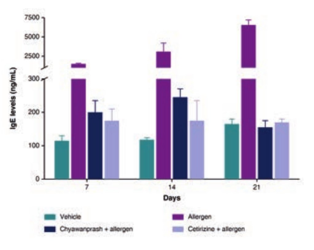 Study 1Estimation of levels of Immunoglobulin E (IgE) in Mice SerumTo evaluate the Anti-allergic potential of Dabur Chyawanprash (DCP) by estimation of levels of Immunoglobulin E (IgE) in mice serum.Study 2Estimation of Levels of Histamine in Plasma of RatsTo evaluate the Anti-allergic potential of Dabur Chyawanprash (DCP) by estimation of levels of Histamine in plasma of rats
Mechanism
- 01Allergen exposure
- 02IgE sensitisation and mast-cell activation
- 03DCP lowers IgE and histamine release
- 04Balanced immune response
Measured readouts
~12-fold
Reduction vs ovalbumin response
~41-fold
Reduction vs ovalbumin response
Near vehicle
DCP-treated animals close to vehicle control
Near baseline
Held across measured time points
26× · 39×
IgE rise at Day 14 and Day 21 (untreated)
~3×
Histamine within 3 minutes (untreated)
Cellular benefits
- Supports a balanced immune response to allergens
- Helps maintain a healthy histamine response
- Supports healthy mast-cell function
- Helps maintain immune balance during seasonal allergen exposure
Interpretation
Acting on both IgE sensitisation and histamine release covers the response before and after mast-cell activation, a wider margin than mediator blockade alone.
Helps maintain a balanced response to seasonal allergens.
DRF in-house animal allergy models.
Estimation of levels of Immunoglobulin E (IgE) in Mice Serum
To evaluate the Anti-allergic potential of Dabur Chyawanprash (DCP) by estimation of levels of Immunoglobulin E (IgE) in mice serum.
Ovalbumin induced allergy in mice
Methodology
The study evaluated the anti-allergic potential of Dabur Chyawanprash (DCP) using an ovalbumin (OVA)-induced allergy model in mice. DCP extract was administered orally at a dose of 1 g/kg, followed by exposure to ovalbumin to induce an allergic immune response. The animals received the initial OVA challenge on Day 0, followed by booster challenges on Days 7 and 14 to reinforce the allergic response and promote sustained immunological sensitization.
To assess the effect of DCP on the allergic response, blood samples were collected on Days 7, 14 and 21, and serum was analyzed for IgE levels. IgE is a key immunoglobulin involved in allergic sensitization and hypersensitivity responses; therefore, changes in serum IgE provide an important indicator of modulation of the allergic immune response.
Cetirizine (10 mg/kg), a commonly used antihistamine with established anti-allergic activity, was included as a positive control to provide a reference for evaluating the anti-allergic effect of DCP.
Overall, this experimental design enabled assessment of whether repeated oral administration of DCP could modulate OVA-induced IgE responses over the course of allergic sensitization, providing in vivo evidence for its potential anti-allergic/immunomodulatory activity.
Data Analysis: Fold increase in IgE levels = {(Conc of IgE in DCP-treated sample – Conc of IgE in Untreated sample)/ Conc of IgE in Untreated sample}*100. Fold decrease in IgE levels = {(Conc of IgE in untreated sample – Conc of IgE in DCP-treated sample)/ Conc of IgE in Untreated sample}*100
Key biomarkers
The biological relevance of this model lies in its ability to assess IgE-mediated allergic sensitization, a key mechanism underlying many allergic responses. Ovalbumin challenge markedly increased serum IgE, indicating activation of the allergic immune pathway. The substantial reduction in IgE following DCP pre-treatment suggests that DCP may modulate allergen-induced immune sensitization and downstream allergic responses.
Since IgE plays an important role in mast-cell activation and release of allergy-associated mediators, suppression of the IgE response may help limit subsequent allergic reactions. Thus, the findings provide in vivo mechanistic evidence for the anti-allergic and immunomodulatory potential of DCP, supporting its potential role in maintaining a balanced immune response to allergens.
Biological pathway
Allergen exposure IgE sensitization mast-cell activation release of allergic mediators allergic response.
Results
Ovalbumin challenge produced a marked elevation in serum IgE levels, confirming successful induction of an allergic immune response. Compared with the vehicle control, serum IgE increased by approximately 26-fold on Day 14 and 39-fold on Day 21, demonstrating progressive strengthening of the allergic response following repeated ovalbumin challenges.
Pre-treatment with DCP substantially suppressed the ovalbumin-induced IgE response. At Day 14, DCP reduced the elevated serum IgE response by approximately 12-fold, while by Day 21 the reduction reached approximately 41-fold relative to the corresponding ovalbumin-induced response. Importantly, DCP-treated animals maintained serum IgE levels close to those observed in the vehicle control group, indicating strong attenuation of the allergen-induced IgE response.
The response observed with DCP was comparable in overall direction to that produced by cetirizine, the reference anti-allergic treatment. These findings suggest that DCP can modulate the IgE-mediated allergic response and reduce allergen-induced immune sensitization in this animal model.
Overall, the results provide in vivo evidence of antiallergic/immunomodulatory activity of DCP, particularly through suppression of the IgE response associated with allergic sensitization. However, because this is an animal model measuring serum IgE, the findings support potential anti-allergic activity rather than directly establishing prevention or treatment of allergic disease in humans.
Scientific interpretation
The data demonstrates a clear IgE-modulating effect of DCP in the ovalbumin-induced allergic model. Ovalbumin challenge produced a marked and progressive elevation in serum IgE levels on Days 7, 14 and 21 compared with the vehicle group, confirming sustained allergen-induced sensitization. In contrast, DCP + allergen treatment substantially reduced the allergen-induced IgE response, with the suppression becoming particularly evident on Days 14 and 21. The DCP response was broadly comparable to the cetirizine + allergen reference group.
Scientific interpretation for consumer benefit claim
The findings provide in vivo mechanistic support for the antiallergic potential of DCP. DCP substantially reduced the allergen-induced rise in serum IgE, indicating that it may help modulate allergic sensitization and maintain a balanced immune response. Since IgE is an important mediator of allergic reactions and can trigger mast-cell activation and release of allergy-associated mediators, reducing the IgE response may help limit the downstream allergic response.
From a consumer-health perspective, these findings support positioning DCP as helping to support healthy immune responses to allergens and maintain immune balance.
Potential consumer benefit claims:
- Supports a balanced immune response to allergens
- Helps support the body’s natural defences against allergic responses
- Supports healthy immune response during allergen exposure
- Helps maintain healthy immune balance
- Supports the body’s natural response to seasonal allergens
- Provides natural immune support against allergen-related challenges
Estimation of Levels of Histamine in Plasma of Rats
To evaluate the Anti-allergic potential of Dabur Chyawanprash (DCP) by estimation of levels of Histamine in plasma of rats
Allergen compound 48/80 induced rats
Methodology
The study evaluated the anti-allergic potential of Dabur Chyawanprash (DCP) using a compound-induced allergic response in rats. Animals were orally administered DCP at a dose of 1 g/kg prior to allergen/compound challenge. The challenge was used to trigger an acute allergic response, enabling assessment of whether DCP could modulate the downstream release of allergy-associated mediators.
Following the challenge, blood samples were collected and plasma histamine levels were quantified. Histamine is a key mediator released during allergic reactions, particularly following activation of mast cells, and contributes to characteristic allergic responses such as vasodilation, increased vascular permeability, itching and inflammatory responses.
Therefore, measurement of plasma histamine provides a biologically relevant endpoint for determining whether DCP can attenuate mediator release following allergen exposure. A reduction in challenge-induced histamine levels in DCP-treated animals would indicate in vivo anti-allergic and mast-cellmodulating activity, providing complementary evidence to the IgE findings from the ovalbumin-induced allergy model.
Data Analysis: Fold increase in Histamine levels = {(Conc of Histamine in DCP-treated sample – Conc of Histamine in Untreated sample)/ Conc of IgE in Untreated sample}*100. Fold decrease in histamine levels = {(Conc of Histamine in untreated sample – Conc of Hitsamine in DCP-treated sample)/ Conc of Hitsamine in Untreated sample}*100
Key biomarkers
The assay is biologically relevant because histamine is one of the major mediators of the immediate allergic response. Following allergen-induced activation of mast cells, histamine is released and contributes to symptoms such as itching, redness, swelling, increased vascular permeability and airway responses.
Therefore, measuring plasma histamine after compound-induced allergy provides a functional readout of downstream allergic mediator release. If DCP reduces the allergen-induced increase in histamine, it suggests that DCP may attenuate mast-cell activation and the subsequent allergic inflammatory response.
Results
The allergen challenge produced a rapid and pronounced increase in plasma histamine, reaching approximately 3-fold above baseline within 3 minutes, confirming a strong acute allergic response and activation of the histamine-mediated pathway.
In contrast, pre-treatment with DCP markedly suppressed the allergen-induced histamine release, maintaining plasma histamine levels close to baseline. The suppression was observed across the measured time points, indicating that DCP was able to attenuate the rapid release of histamine following allergen exposure rather than merely delaying the response.
Biologically, these findings suggest that DCP may modulate mast-cell activation and subsequent histamine release, an important downstream event in immediate hypersensitivity reactions. Reduced histamine availability could potentially limit several physiological manifestations of allergic responses, including increased vascular permeability, tissue swelling, itching and inflammatory responses.
Scientific interpretation
The findings demonstrate that DCP significantly attenuated the acute allergen-induced histamine response. Allergen challenge caused a rapid increase in plasma histamine, reaching approximately 3-fold above baseline within 3 minutes, confirming activation of the immediate allergic response. Pre-treatment with DCP maintained histamine levels close to baseline and significantly suppressed histamine release across the evaluated time points.
Scientific interpretation for consumer benefit claim
The findings provide in vivo mechanistic support for the antiallergic potential of DCP. The rapid allergen-induced increase in plasma histamine was markedly suppressed by DCP pre- treatment, with histamine levels maintained close to baseline. Since histamine is a key mediator of immediate allergic responses, this suggests that DCP may help modulate mast-cell activation and excessive histamine release, thereby supporting a more balanced response to allergen exposure.
From a consumer-benefit perspective, these findings support positioning DCP as helping to maintain healthy immune responses during allergen exposure and providing natural support against allergy-associated immune reactions. When combined with the IgE data, the evidence provides complementary support for an anti-allergic immune-balance mechanism.
Potential consumer benefit claims:
- Supports a balanced immune response to allergens
- Helps support the body’s natural defence against allergens
- Supports healthy immune responses during allergen exposure
- Helps maintain a healthy histamine response
- Supports healthy mast-cell function
- Helps maintain immune balance during seasonal allergen exposure
- Provides natural anti-allergic immune support